NEUR0010 Neurobiology of Brain Injury and Disease (NEUR0010)
Hochschule
University College London (UCL)
Complete notes and all additional reading for the module including textbook notes, assigned reading, and extended reading. Evidence and research papers allocated in tables and broken down into background, aims, methods, results, conclusions, and critical thinking. Broken down into chapters and subc...
NEUR0010 Neurobiology of Brain Injury and Disease (NEUR0010)
Alle Dokumente für dieses Fach (17)
1
bewertung
von: andrijadjuric612 • 4 Monate vor
Verkäufer
Folgen
denh11323
Deine Reviews
Inhaltsvorschau
NEUR0010
NEUROBIOLOGY OF BRAIN INJURY AND DISEASE
Maria Thom: introduction to neuropathology
KEY POINTS
1. Role of neuropathology
2. Processing and handling of brain tissue for examination of any neuropathology
3. Stains and immunohistochemistry to visualize cell types
4. Range of cytopathology and tissue reactions that give clues as to disease processes and further tests
TISSUE HANDLING PATHWAYS
1. Samples gathered
a. Neurosurgical specimens
b. CSF cytology
c. Nerve and muscle biopsies
2. Samples sent to the lab
3. Tissue samples frozen
a. Formalin fixed
b. Processed for histology/light microscopy
c. Cytology preparations for fluid
4. Diagnosis OR
a. Indications of nature of disease process
5. Direct further molecular, genetic, other clinical investigations
6. Discussed in a multidisciplinary team meeting
7. Appropriate clincal treatment/management
TISSUE PROCESSING AND STAINING
Fixation (e.g. formalin)
- Cross links proteins to prevent enzyme digestion (autolysis)
- Tissue embedded in paraffin wax to enable thin sections to be cut
MAIN TYPES OF NEUROPATHOLOGY SPECIMENTS
1. Surgical speciments
a. Lobectomies
b. Tumour resections
c. Brain biopsies
2. Cytology samples (cells only)
a. Intraoperative smears
b. CSF cytology → to check the nature of lesion and if the biopsy of the lesion is on target
i. Needle biopsy
ii. Fresh sample in laboratory
iii. Small piece smeared, fixed and stained with H&E
1. Brain does not have any collagen (how does this allow for smearing?)
iv. Diagnosis reported back to theatre within 20 mins
3. Postmortem specimens
, a. Whole brain retention at postmortem: tissue fixation in formaldehyde for 2-3 wweeks to then
examine brian
b. Fix brain for a shroter interval, examin, sample small regions for histopathology and return brain to
the body
c. Photography of fresh macroscopic brain at postmortem but only smsall samples of tissue are kept
d. Value of postmortem examinations
i. Clinical audit/epidemiology
ii. Investigations in sudden deaths
iii. Identification and understanding of new disease e.g. variant CJD in 1990s, COVID19
iv. Brain banking for research into neurological disease to identify pathomechanisms and new
treatments
STRATEGIC TISSUE SAMPLING FOR HISTOLOGY
level 1: fornix
level 2: M. body
level 3: LGN
- Small tissue blocks are taken from specific regions
- Regions sampled will depend on
o Clinical diagnosis
o Neuropathology findings
- For many conditions there are standardized sampling protocols
Hippocampus
- Hypoxic ischemic damage
- Neurodegeneration and aging – memory loss
- Seizures and epilepsy
KEY MARKERS WHEN ASSESSING BRAIN HISTOPATHOLOGY
1. Do the neurons and glial cells appear normal?
a. In distribution
b. Relative number – loss or increase
c. Abnormal material
2. Is there any inflammation or infiltrate in the tissues?
3. Do the myelin and axons appear normal?
a. Loss or breakdown
4. Do the neuropil appear normal?
a. Plaques or aggregates
5. Do the cerebral blood vessels appear normal?
,HISTOLOGY STAINS
1. Tinctorial stains (vegetable dyes)
a. Traditional stains such as Haematoxylin and Eosin (H&E)
2. Luxol fast blue stain (myelin) with Cresyl violet (Nissl stain for neurons)
IMMUNOHISTOCHEMISTRY
Introduced in 1980s and now routinely used in research and diagnostics
Diagnostics and markers → brown chromagen (DAB) to detect cells and multi-channel labelling with combos of
fluorescently tagged antibodies
- Delineate cell types/lineages (astrocytes)
- Mutated cells in tumour diagnosis (tumours with IDH mutation)
- Cell division/proliferation (with Ki67 marker)
- Abnormal protein accumulation (tau in AD)
- Cell receptors (serotonin on neurons)
- Viral proteins (JC virus in PML)
Visualising cortical layers
NEURONE MORPHOLOGY
1. Prominent nucleolus
2. Prominent ReR (Nissl bodies)
3. Pigments
4. Lipofuscin: age pigment in adult neuorns → biproduct of membrane turnover
5. Neuromelanin 0 in catecholaminergic neurons
6. Cytoskeleton
a. Microtubules
b. Neurofilaments
c. Microfilaments
,BASIC CELLULAR REACTIONS TO CNS INJURY
NEURONAL LESIONS
- Examination after injury with H&E = eosinophilia of cytoplasm + shrinkage and hyperchromasia of nucleus +
disappearance of nucleolus
- Subsequent to disintegration of cell → neuronophagia by scavenger cells
- Reduction of cell bodies in the area compared to normal (>30% of normal population)
NERVE CELL ATROPHY
What? Wide range of irreversible neuronal injury causing slowly-evolving death of cell
Morphological characterization:
- Retraction of cell body
- Diffuse basophilia of cytoplasm
- Pyknosis and hyperchromasia of nucleus of neuron in absence of inflammatory reaction
When?
- Degenerative diseorders involving interconnected neuronal systems
- Anterograde and retrograde transsynaptic degeneration
o Can occur in lateral geniculate body after optic nerve lesion
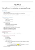
Figure 1: In one neuron, on the left, only the nucleus is stained, whereas in the other, which is at a later stage of the programmed
cell death process, the entire cell body is stained. Compared to a normal neuron, on the right, both apoptotic neurons have
similar morphologic features and show pyknotic nucleus and shrunken cytoplasm
Apoptotic neurons will show positive in situ end labelling of internucleosomal DNA fragmentation or be demonstrated
by activated caspase-3 immunostaining
ACUTE NEURONAL NECROSIS
What? Cell death that occurs in acute injuries (e.g., anoxia and ischemia) or in acute pathological processes
(hypoglycemia or exposure to excess excitotoxic NTs)
- Cytoplasmic organelels and cell membrane ruptures → cell death
Evidence
Postmortem + light and electron microscopy shows the following changes from 12-24 hours after insult
1. Cytoplasmic microvacuolation caused by swelling of mitochondria and ER
2. Shrinkage of cell body with retraction of cellular outlines
3. Disappearance of Nissl bodies with eosinophilic condensation of cytoplasm
4. Condensation of nuclear chromatin and nuclear puknosis
5. Late disappearance of nuclear chromatin causing increased acidophilia of nucleus (merges into surrounding
cytoplasm)
Mineralization/ferrugination: occurs when dead neurons become encrusted with basophilic mineral deposits (Fe and
Ca)
CENTRAL CHROMATOLYSIS
Morphological characterization
- Swelling of cell body
- Disappearance of Nissl bodies
- Flattening and eccentric displalcement of nucleus to the periphery
Where: lower motor neurons → represents reparative reaction of cell body to a lesion of the axon
NEURONAL INJURY
Limited capacity to survive injury → Post-mitotic cells
Common forms of neuronal injury
- Hypoxia
- Direct trauma
- Infection
- Seizure activity
- Metabolic causes
Neuronal cell death mechanisms
- Excitotoxic neuronal injury
- Oxidative stress – reactive oxygen species
- Mitochondrial dysfunction
- Protein aggregation/inclusions in cell causing dysfunction
- Inflammatory pathway activation
Mode of neuronal death
,Dependent on underlying cause
- Induction of regulated cell death pathways
- Intrinsic and extrinsic apoptosis pathways
- Necroptosis
- Pyroptosis
- Autophagy
- Ferroptosis
NEURONAL INCLUSIONS
Can be cytoplasmic or nuclear and can indicate:
- Infection
- Abnormal accumulation of protein
- Metabolic disorder
- Genetic disorder
- Age related and no pathological significance
Basophilic flame shaped inclusion
Hirano bodies: brightly eosinophilic inclusions of varied shape. Majority are small rod-shaped structures with rouned
ends → seen in hippocampus in ageing
Spherical eosinophilic intracytoplasmic inclusion
- Lewy bodies in PD in substantia nigra
,Rosenthal fibres
- Coarse astroglial prcesses
- Observed in astrocytic tumours
- Observed in alexander’s disease (mutations in GFAP →
leucodystrophy)
AMYLOID PROTEIN
- Extracellular, proteinaceous deposit exhibiting beta sheet structure
- Identified by apple-green birefringence when stained with congo
red and seen under polarized light
- Accumulated in the brain with ageing
- In blood vessels and parenchyma as plaques seen I and
- Amyloid in blood vessels predisposes to cerebral haemorrhage and can be sporadic or genetically inherited
ASTROCYTES
- Outnumbers neurons ~4:1
- Old notion was they were just supporting CNS cells
Known Functions
- Contribute to CNS homeostasis (glutamate/K+/adenosine)
- BBB function (end feet make contact with blood vessels in neurons)
- Release cytokines (pro-inflammatory), growth factors and gliotransmitters
- Proliferate in response to injury → form glial scar (gliosis) to limit or localize damage
- Progenitor cells source (tumourgenesis)
- Useful as a sensitive biomarker for brain disease
- Not specific pathology finding as gliosis is a universal reaction to many disease processes
GFAP: glial fibrillary acidic protein
- Intermediate filament specific to astrocutes to visualize cells
,OLIGODENDROCYTES
- Round nuclei particularly visible in white matter
- Can be seen on H&E or luxol fast blue stain
- Provide myelination to axons
- Oligodendrocytes derived from oligodendolial progenitor cells (OPC)
o OPC (NG2) cells
▪ Present in adult brain
▪ Important in maintenance and repair of myelin
▪ Immunomarkers used to show maturation
▪
▪
,MICROGLIAL CELLS
- CNS macrophage network
- Can be activated or nonactivated
-
Macrophage activation states and immunophenotyping
, BLOOD VESSELS AND BBB
MTOR PATHWAY MUTATIONS IN FCD
MTOR Pathway
- Promotes cell growth and proliferation over differentiation
- GERMLINE mutations in TSC1 and 2 genes in this pathway in TB
- Activation of mTOR pathway in FCD using immunohistochemistry 2006
- SOMATIC mutations in abnormal cell types (Dysmorphic neurons and glia) identified with deep sequencing in
FCD cases in 2015
Potential New Treatment Targets: Rapamycin and other drugs that target the mTOR pathway as possible alternatives
to surgery
NEURODEGENERATIVE DISEASE
Definition
A heterogeneous group of disorders that are characterized by the progressive degeneration of the structure and
function of the central nervous system or peripheral nervous system.
Commonest = AD and PD
Common Pathomechanisms Underlying Neurodegenerative Disease
1. Neuronal protein misfolding and accumulation/neuronal dysfunction/cell death
a. Beta amyloid (AD)
b. Tau (AD, CTE, etc)
c. Prion proteins (CJD)
d. Alpha-synuclein (PD, MSA)
e. TDP-43 (LATE)
2. Impaired protein degradation pathways
a. Ubiquitin pathway
Alle Vorteile der Zusammenfassungen von Stuvia auf einen Blick:
Garantiert gute Qualität durch Reviews
Stuvia Verkäufer haben mehr als 700.000 Zusammenfassungen beurteilt. Deshalb weißt du dass du das beste Dokument kaufst.
Schnell und einfach kaufen
Man bezahlt schnell und einfach mit iDeal, Kreditkarte oder Stuvia-Kredit für die Zusammenfassungen. Man braucht keine Mitgliedschaft.
Konzentration auf den Kern der Sache
Deine Mitstudenten schreiben die Zusammenfassungen. Deshalb enthalten die Zusammenfassungen immer aktuelle, zuverlässige und up-to-date Informationen. Damit kommst du schnell zum Kern der Sache.
Häufig gestellte Fragen
Was bekomme ich, wenn ich dieses Dokument kaufe?
Du erhältst eine PDF-Datei, die sofort nach dem Kauf verfügbar ist. Das gekaufte Dokument ist jederzeit, überall und unbegrenzt über dein Profil zugänglich.
Zufriedenheitsgarantie: Wie funktioniert das?
Unsere Zufriedenheitsgarantie sorgt dafür, dass du immer eine Lernunterlage findest, die zu dir passt. Du füllst ein Formular aus und unser Kundendienstteam kümmert sich um den Rest.
Wem kaufe ich diese Zusammenfassung ab?
Stuvia ist ein Marktplatz, du kaufst dieses Dokument also nicht von uns, sondern vom Verkäufer denh11323. Stuvia erleichtert die Zahlung an den Verkäufer.
Werde ich an ein Abonnement gebunden sein?
Nein, du kaufst diese Zusammenfassung nur für 24,86 €. Du bist nach deinem Kauf an nichts gebunden.