Summary of Human development, biomedical sciences 1st year
68 mal angesehen 7 mal verkauft
Kurs
Human Development (AB_1140)
Hochschule
Vrije Universiteit Amsterdam (VU)
A summary of the course human development, teached by S. Spijker in the first year of biomedical sciences. Contains all lecture notes and exam material.
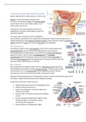
Lecture 2a male reproductive system
Martini H28, H29 (1177-1185), Larsen p.1-13 H1 (1-24)
Gonads = organs that produces gametes and
hormones, transported by ducts, the accessory glands
secrete fluids into the ducts. Males produce 1 or 2
million sperm cell per day.
Important for the male reproductive system are:
hypothalamus, pituitary, adrenal glands, testes and
reproductive glands.
The sperm makes a long way: testis → epididymis,
ductus deferens, ejaculatory duct. Spermatozoa development requires lower temperatures, to
regulate temperature the cremaster and dartos muscles contract. Semen is made in seminiferous
tubules between the septa, cells travel via rete testis to the epididymis.
Spermatogenesis
Seminiferous tubules contain spermatogonia (=stem cells for spermatozoa) and
sustentacular cells (=sustain and promote development of spermatozoa).
Spermatogonia goes through mitosis, one of them forms primary spermatocyte
going into meiosis 1 (other remains spermatogonium)→ 2 secondary
spermatocytes formed → going into meiosis 2 forming 4 haploid spermatids →
undergoing spermiogenesis forming spermatozoa → loosing connection with wall
and enter lumen. (whole process in 9 weeks).
Spermiogenesis
Most organelles and cytoplasm needs to be lost. Sustentacular cells phagocytose
shed cytoplasm and provide nutrients. Acrosomal vesicle = fused saccules of Golgi
and peroxisome to form acrosomal cap (contains degrading enzymes). Mature
spermatozoon only has mitochondria and DNA, making sperm faster.
Anatomy of the spermatozoon has 4 compartments: head (= nucleus and acrosomal cap), neck (=
centrioles), middle (=mitochondria) and tail (=flagellum moving).
Sustentacular cells have 6 major functions:
• Maintain blood-testis barrier → having tight junction
• Support mitosis and meiosis →
• Support spermiogenesis → promote division by
chemical stimuli ABP
• Secrete inhibin → female and male, gives feedback
control
• Secrete androgen-Binding Protein (ABP) → male,
binding androgens in seminiferous tubule,
stimulating spermatogenesis
• Secrete Mullerian-inhibiting factor (MIF)
,Hormonal regulation
GnRH from hypothalamus stimulates anterior lobe pituitary
gland → secretion LH and FSH.
FSH stimulates sustentacular cells to make ABP and
spermatogenesis → secretion inhibin giving negative
feedback FSH
LH causes secretion testosterone via interstitial cells
promoting male sexual characteristics, inhibiting GnRH.
Maturation
Testes produce physically mature spermatozoa but they can
not fertilize oocyte. They are incapable of locomotion,
moved by cilia in efferent ductulus (epididymis) functions:
• Monitors / adjusts fluid produced by seminiferous
tubules
• Recycles damaged spermatozoa
• Stores/protects spermatozoa until use
Leaving the epididymis still immobile, to become motile
have to undergo capacitation: 1. Mixing with secretion
seminal vesicles 2. Exposure to female reproductive tract, destabilization of acrosome due to
proteolytic and glycosidase enzymes removal of steroids.
Capacitation
In uterus sperm induces host reaction, leucocytes engulf spermatozoa. Isthmus functions as sperm
store. Non-identified chemotactic cues regulate increase in calcium: attached sperm have low
calcium concentration (maintain longevity and function), detached sperms have high calcium
concentration (enables ore vigorous motility).
Loss of membrane cholesterol is vital to
fertilize egg, during passage to uterus
albumin and high-density lipoproteins act as
cholesterol acceptors. GPI-anchored
proteins are inhibitors of sperm-egg binding
are lost from membrane. ACE cleaves GPI-
anchored proteins form sperm membrane
→ testis-specific isozyme of ACE is only in
developing spermatids.
Sperm
Male glands have 4 major functions: activating spermatozoan providing nutrients, propelling along
reproductive tracts and producing buffers.
Seminal vesicles = activate secretory gland and contain fructose, prostaglandins and fibrinogen.
Prostate gland = secrete acidic prostate fluids. Bulbourethral glands = secrete alkaline mucus
working as lubricant.
,Lecture 2b Female reproductive system
Martini H28, Larsen h1 (25-33)
Differences between male and female are that in female
from one oocyte, only 1 egg cell is formed. Female
reproductive system has 2 functions: produce sex
hormones/gametes, protect developing embryo and nourish
infant. Ovaries have 3 main functions:
• Production immature female gametes = oocytes,
produced in cortex of ovary in a follicle.
• Secretion of female sex hormones = estrogens and
progestins → feedback uterine cycle
• Secretion of inhibin → feedback control pituitary FSH
Oocytes are produced in cortex of the ovary in a follicle.
Oogenesis
Mitosis of oogonia takes place before birth. Oogonia divide by mitoses, only
1 of cells becomes primary oocyte. Meiosis starts at the menarche and ends
at menopause. Ovulation = end meiosis 1 and start meiosis 2.
Gonad formation
Primordial germ cells migrate to genital ridge in developing embryo (4-6
weeks), in oogonia after this mitosis starts (6 weeks-3months). All cells are
suspended in prophase of meiosis 1 = diplotene stage. Many of those
degenerate, only ones near cortex survive.
Meiosis
Primary oocytes and surrounding flat epithelial cells = primordial follicle. At
puberty each month a set of primordial follicles undergo maturation →
become antral (=forming cavity). Follicle cells in outside = thecal cells, on
inside surrounding oocyte = granulosa cells (travel along with oocyte upon
ovulation). Also forming layer of glycoprotein (inside) = zona pellucida and
layer granulosa (outside) = corona radiata.
2 characteristics of meiosis:
1. Cytoplasm of primary oocyte divides unevenly (asymmetric cytokinesis) → producing 1
ovum and 1-3 polar bodies
2. Ovary releases secondary oocyte (no mature ovum)→ suspended metaphase meiosis 2, only
completed upon fertilization
Meiosis 1 is started by raising FSH, each month ovarian cycle produces 1 secondary oocyte. Viability
of ovulated oocytes declines over time.
3 times a pause: diplotene stage, metaphase meiosis 1 and metaphase meiosis 2. These pauses are
done by protein complexes. Phosphorylation by kinases: MPF and dephosphorylation by
phosphatases: calcineurin. Second messenger cascades involving cAMP/cGMP activate kinases.
, in diplotene stage increased NO signaling increased
cGMP, thecal and granulosa cells release purines →
increased cAMP levels (PDE3A inactivation) via PKA
activation and inhibition MPF.
MPF = M-phase promoting factor, breakdown of MPF
to go to next phase.
Ovarian cycle
Ovarian cycle has follicular phase (=preovulatory) and
luteal phase (=postovulatory).
Follicular phase
Prophase of meiosis 1 and at end metaphase meiosis
1 → LH peak. From primordial follicle in egg nest →
primary follicle → secondary follicle → tertiary follicle.
After this, LH surge finishes meiosis 1 to generate one
secondary oocyte and a polar body. Only secondary
oocyte enters meiosis 2.
Luteal phase
Starts after ovulation formation of corpus luteum by
LH, which produces progesterone, no fertilization → degeneration after 12 days.
Uterine cycle
Few hours before ovulation, start ciliary movement and peristaltic contraction. 3-4 days from
infundibulum to uterine cavity. Fertilization during first 12-24 hours. Uterus itself is muscular organ,
giving mechanical protection, nutritional support and waste removal. It is supported by broad
ligament and 2 pairs suspensory ligaments. Unterine wall is made of
3 layers:
• endometrium = thin, inner glandular mucosa,
o basilar zone
o functional zone degrades during the menstruation
• myometrium = outer muscular layer
• perimetrium = incomplete serosa continuous with
peritoneum
hormonal control
pulse frequency GnRH depends on ovarian cycle, cells are sensitive
to different pule frequencies. FSH stimulates follicles, inhibin and
estrogens which stimulate LH → stimulates secretion progesterone
by corpus luteum → stimulates endometrial growth.
Body temperature
Luteal phase → progesterone. Follicular phase → estrogen, lower
body temperature. Upon ovulation a larger dip, the day after
coming back to normal.
Alle Vorteile der Zusammenfassungen von Stuvia auf einen Blick:
Garantiert gute Qualität durch Reviews
Stuvia Verkäufer haben mehr als 700.000 Zusammenfassungen beurteilt. Deshalb weißt du dass du das beste Dokument kaufst.
Schnell und einfach kaufen
Man bezahlt schnell und einfach mit iDeal, Kreditkarte oder Stuvia-Kredit für die Zusammenfassungen. Man braucht keine Mitgliedschaft.
Konzentration auf den Kern der Sache
Deine Mitstudenten schreiben die Zusammenfassungen. Deshalb enthalten die Zusammenfassungen immer aktuelle, zuverlässige und up-to-date Informationen. Damit kommst du schnell zum Kern der Sache.
Häufig gestellte Fragen
Was bekomme ich, wenn ich dieses Dokument kaufe?
Du erhältst eine PDF-Datei, die sofort nach dem Kauf verfügbar ist. Das gekaufte Dokument ist jederzeit, überall und unbegrenzt über dein Profil zugänglich.
Zufriedenheitsgarantie: Wie funktioniert das?
Unsere Zufriedenheitsgarantie sorgt dafür, dass du immer eine Lernunterlage findest, die zu dir passt. Du füllst ein Formular aus und unser Kundendienstteam kümmert sich um den Rest.
Wem kaufe ich diese Zusammenfassung ab?
Stuvia ist ein Marktplatz, du kaufst dieses Dokument also nicht von uns, sondern vom Verkäufer lente90. Stuvia erleichtert die Zahlung an den Verkäufer.
Werde ich an ein Abonnement gebunden sein?
Nein, du kaufst diese Zusammenfassung nur für 6,99 €. Du bist nach deinem Kauf an nichts gebunden.