This is a summary I made while following the Bacterial Pathogenesis course. In the document, I have also included a summary of the pre-requisite knowledge videos/papers they provided to follow the course.
Review prerequisite knowledge
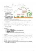
Biofilm formation
1. Specific environmental
signal genetic change to
free living planktonic cells
2. Attach to nearby surfaces by
means of LPS, pili, flagellum
3. Bacteria coat the surface
with polysaccharide or
glycoprotein, more
planktonic cells can attach
there
4. Get rid of flagella, move via
twitching motility (extension
and retraction of pilus)
5. More and more cells bind, they form microcolonies
6. Send chemical signals (quorum sensing) to communicate with each other (continually made by
individual cells)
7. Trigger genetic changes to bind to other cells and to the substrate. Leads to more production of the
matrix
8. ECM protects the cells from outside threats (e.g., antibiotic resistance)
9. Can form channels for nutrient and water flow, biofilms form in a rich nutrient environment
10. When nutrient is low, they can remove the ECM and revert to their flagellated planktonic form and
find a better location
Advantages:
- Remain at a suitable location
- Cells Are In Close Proximity, quorum sensing, and DNA transfer
- Collaboration, task division
- Protection–against predation, immune system and antimicrobials.
Chemotaxis
Flagella can rotate clockwise spin into a tumble or counterclockwise spin into a run. Chemoattractants
are molecules that can influence bacterial movement into a certain direction for example food and urea
(in the case of H. pylori). It involves several protein key players:
1. Methyl accepting chemotaxis protein (MCP) → Interacts with CheA and CheW, receptor of the
chemoattractant
2. CheA → Auto-phosphorylates and donates phosphates to CheY
3. CheW → Assists interaction of CheA and MCP
4. CheY → Response regulator. Phosphorylated by CheA and dephosphorylated by CheZ. Determines
which rotation direction the flagella go.
,5. CheB → Response regulator. Demethylates MCP.
6. CheZ → Dephosphorylates CheY
7. CheR → Methylates MCP
Without a chemoattractant, CheA auto phosphorylates itself using ATP. It then donates phosphates to
CheY which moves to the flagella and induces a clockwise rotation (tumble since no chemoattractant is
bound to MRP). CheZ then dephosphorylates CheY. CheR slowly methylates MCP and CheB
(phosphorylated by CheA) removes the phosphate groups on MCP at a faster rate.
When a chemoattractant is bound to MRP, it decreases the rate of auto-phosphorylation of CheA which
subsequently decreases phosphorylation of CheY and CheB. CheR methylation dominates CheB.
Furthermore, phosphorylated CheY goes to the flagellum and causes it to run by rotating
counterclockwise.
Flagella structure
The flagella is made out of 3 main parts: basal body
(intracellular), the flexible hook (extracellular, outside
the cell wall), and the filament. The basal body
consists of a rod and rings. The rings are embedded in
the inner membrane, peptidoglycan (stator), and
outer membrane. The stator is embedded in the
peptidoglycan as this is a rigid layer, holding this
stationary unit in place (can also be in the outer
membrane in gram negative).
The rotor portion uses a proton gradient from the
outer membrane. Protons travel across via MotA and
MotB, parts of the stator. Each stator molecule has 2
MotB and 2 MotA subunits:
1. H+ bind to the aspartic acid residues in MotB
2. Conformational change in MotA → 1st power stroke
3. Proton enters the cytosol
4. Loss of proton leads to conformational change of MotA → 2nd power stroke
Assembly:
1. 26 subunits of FliF come together in the plasma membrane → MS ring
2. FliG along with FliM and FliN proteins make up the rotor under MS ring → C ring
3. Flagella associated proteins are transported through this ring from the bottom to the top
4. MotA and MotB form the stationary part, both integral proteins but MotB is also anchored to
peptidoglycan
5. Rod proteins move up through the ring and form proximal to distal, assisted by cap proteins
(pentameric complex where the subunits of the flagella are attached to this frame)
,6. L and P ring form in gram negative, in the outer membrane forming a bearing
7. Cap protein exposed out of the cell membrane, dissociates, replaced by hook cap that guides the
assembly of the hook protein (flexible and strong, can change angles of the protein subunits to bend
the flagella around)
8. After hook is assembled, hook cap dissociates, and a bunch of junction proteins assemble between
hook and future filament
9. Filament cap is put on and filament proteins (flagellin) travel from the hollow rod proteins to form
the distal ends
10. The filament cap is also rotating while the filament is being formed, allowing it to stack in a helical
fashion
Quorum sensing
Vibrio fischeri communicates with each other. In low cell numbers, nothing happens, but when there is a
certain amount, they all suddenly emit light. Individually, they produce molecules which is a light
emitting signal, but it is in low concentration. When a lot of them start producing more molecules such
that it outnumbers the cell population and when the molecule reaches a certain amount, they all signal
for bioluminescence.
These bacteria have symbiosis with the bobtail squid. The squid has a detector on the back and a
shutter on top of 2 light organs on its bottom, housing these bacteria. The squid senses how much
moon/star light it has outside and adjusts the shutter to match the amount of light such that it doesn’t
make a shadow. This is beneficial to hunt and cannot get detected by predators. When the sun comes
up, the squid buries itself in the sand and pumps out 95% of the bacteria. As the day goes by, the
bacteria doubles and releases the molecule and lights up at night.
The molecules are all related in all bacteria species. There is intraspecies specificity of the molecules.
Each bacteria uses their own molecules to count their own population. Once they have enough
population, they all start secreting these molecules in a high amount such that it starts the virulence.
They also have interspecies communication. So, they know how much of themselves and how much of
the other populations. The universal molecule is produced by every single bacterium by the same
enzyme.
We can target this communication system (makes them mute and deaf) by targeting the intraspecies
molecule (jamming receptor by using an antagonist, species specific) or by making an antagonist of the
interspecies communication which can be used as a broad spectrum antibiotic for all bacteria. We can
also use analogs for good bacteria to increase their activity vs pathogens.
When a bacterium enters a host, it doesn’t immediately secrete its virulence. They make these
communication molecules to know how many there are, and once they have enough population, they
then generate their virulence.
, Mucosal defense
There are different components of the intestinal mucosal defense. Apart from the immune cells (DCs,
Macrophages, B cells, T cells), there are mucus (synthesized by Goblet cells), IgA antibodies (Bind
invasive bacteria), and Antimicrobial peptides (synthesized by Paneth cells).
The intestinal mucus layer
In the healthy state, the outer mucus
layer is in contact with commensals.
Commensals are separated with the
epithelial barrier by the mucosal system
which leads to anti-inflammatory
signaling. Furthermore, epithelial cells
express transmembrane mucins. In a
diseased condition, bacteria is no longer
separated from the epithelial cells.
These bacteria are capable of moving
through the mucus layer, invade
epithelial cells, and spread through the
body by means of systemic infections.
The soluble mucus layer is the first line of defense. It also has different portions depending on the
organs and location. Throughout the stomach to the end of the colon, there are different properties of
the secreted mucus layer.
- Stomach: there are 2 layers (inner and outer) mucus layers maintained
- Small intestine: the inner mucus layer is virtually absent. Outer mucus layer more prominent to
allow migration of nutrients through this layer
- Large intestine: thick outer mucus layer especially in the colon. The colon is the site that harbors
the largest microbiota, this thick layer is used to separate them from the epithelial. Mucus layer
feeds the microbiota.
The composition and function of mucus layer is shown below:
Glycosylation of mucin
There are many different sugar sources available in the small intestines which are used to form glycans.
These glycans are attached to mucins, which makes them highly glycosylated. Microbiota feed on the
glycans, the most important ones are sialic acid (SA) and fucose. They can cleave these sugars to feed
on. For commensals, the mucus layer is a physical barrier as well as a source for nutrients.
Alle Vorteile der Zusammenfassungen von Stuvia auf einen Blick:
Garantiert gute Qualität durch Reviews
Stuvia Verkäufer haben mehr als 700.000 Zusammenfassungen beurteilt. Deshalb weißt du dass du das beste Dokument kaufst.
Schnell und einfach kaufen
Man bezahlt schnell und einfach mit iDeal, Kreditkarte oder Stuvia-Kredit für die Zusammenfassungen. Man braucht keine Mitgliedschaft.
Konzentration auf den Kern der Sache
Deine Mitstudenten schreiben die Zusammenfassungen. Deshalb enthalten die Zusammenfassungen immer aktuelle, zuverlässige und up-to-date Informationen. Damit kommst du schnell zum Kern der Sache.
Häufig gestellte Fragen
Was bekomme ich, wenn ich dieses Dokument kaufe?
Du erhältst eine PDF-Datei, die sofort nach dem Kauf verfügbar ist. Das gekaufte Dokument ist jederzeit, überall und unbegrenzt über dein Profil zugänglich.
Zufriedenheitsgarantie: Wie funktioniert das?
Unsere Zufriedenheitsgarantie sorgt dafür, dass du immer eine Lernunterlage findest, die zu dir passt. Du füllst ein Formular aus und unser Kundendienstteam kümmert sich um den Rest.
Wem kaufe ich diese Zusammenfassung ab?
Stuvia ist ein Marktplatz, du kaufst dieses Dokument also nicht von uns, sondern vom Verkäufer feliciaamelie. Stuvia erleichtert die Zahlung an den Verkäufer.
Werde ich an ein Abonnement gebunden sein?
Nein, du kaufst diese Zusammenfassung nur für 7,49 €. Du bist nach deinem Kauf an nichts gebunden.