8TM00 Bone structure and function
Lecture 1 – Basics & clinical issues
BASICS
Different levels of organization
• Femur is biggest bone
• In center it consists of dense material that forms hollow tube, called cortical/compact bone
o Has some porosity, but very small (too small for naked eye)
• Near end it is more filled with porous type of bone, called
trabecular/cancellous/spongy bone
o Consists of struts of bone material called trabeculae
o Spacing ~1 mm
Trabecular bone
• Longitudinal organization in trabeculae is seen
• This is due to platelet like structures in trabeculae that are laid down by
cells in direction of trabeculae
• In tissue you have osteocytes located in small spaces called lacunae
within bone tissue.
• Osteocytes have a lot of process, they connect to other osteocytes.
o Processes are laid down in canals, called canaliculi.
Compact bone
• Cylindrical structures called osteons
• At center of osteon you have cavity, the central canal. Here you
have nerves and blood vessels.
• Osteocytes cells are present within an osteon, here they are in
their lacuna and connected via canaliculi (similar to what is seen
in trabecular bone).
Different length scales
• At meter, largest bones in body such as femur. So organ level
• At millimeters, you look at cancellous or cortical bone regions.
• At micrometers, at length scale where you recognize cells (~10 μm)
• At nanometers, actual tissue material in cancellous and cortical bone is more or less the same and
consists of collagen and hydroxyapatite.
Focus is on mechanics: why?
Mechanical loading plays an important role during bone
• Formation
• Growth
• Adaptation
• Functioning
1
,Formation & development
Formation
• At embryonal stage, bone is nothing more than cartilage plug
• Cartilage plug starts to mineralize at center at some point
• Mineralization extends plug gets bigger, at some point vascularization happens turns in real
bone tissue
• Now you have bone tissue with cartilage plugs at the
ends
• When bone further grows you get secondary
mineralization zone in cartilage ends that are left,
this gets penetrated by vessels and then turns into
real bone.
• In center you have bone, cartilage between bone
parts become growth plate and outer cartilage
layers turn into the joint surface.
Growth
• Growth plate consists of hypertrophic chondrocytes
• As hypertrophy of the chondrocytes occur, the bone on top is
being pushed away further outwards bone grows
• After hypertrophy, they mineralize the surroundings.
• Mineralized cartilage is turned into real bone
• Around age of 17 growth plate is closed and growth stops
Adaption
• Bones are not yet inert, they can still adapt.
• Trabecular architecture can adapt, e.g. if you load your bone more it becomes more dense.
• Can also be mis adaption, resulting in joint/bone diseases
During all these stages, mechanical factors play an important role.
Formation
• From stem cells tissue can develop
• Depending on strain and hydrostatic pressure different types
of tissue can develop
• Mainly hydrostatic pressure tissue becomes cartilage
• Mainly strain fibrous connective tissue
Development
• Bone structure of tibia in pig: trabeculae
o At 6 weeks disorganized
o Over time structure organizes itself
o Becomes more vertically oriented
• Human femur
o Trabeculae organization is not random
o Preferred orientation in certain direction at
different locations due to mechanical loading
2
,Remodeling & adaption
• Wolff’s law: mass and orientation of trabecular structure are adapted to its mechanical load.
• If a bone fractures it might grow back together and form new optimized architecture structure.
• Bone tissue is constantly renewing itself: bone turnover
• Micro-cracks are repaired
• Enables adaptation of the structure
Trabecular bone
• Osteoclasts reserve bone tissue, can dig cavity in bone tissue
• Osteoblasts: deposit bone tissue in this cavity
• By doing so you get new bone tissue
Cortical bone
• Osteoclasts dig tunnel in direction of longitudinal axis of bone
• Osteoblasts follow osteoclasts and fill tunnel again, but leave small
center that can be filled by blood vessels and nerves.
CLINICAL ISSUES
Skeletal deformities
• Can be due to misalignment of force
• Mechanical loading plays an important role during growth
• A growth plate normally aligns perpendicularly to the force that is
acting on the bone, so if force is on different angle, then growth plate is at different angle which
results in growth of bone in different direction.
• Aside from orientation, the magnitude of force is also important for bone development. If there
is very little force, then not a lot of bone development.
• Idiopathic deformities: even when orientation and magnitude is normal, you can get skeletal
deformities. An example is scoliotic spine, the vertebrae deform force not straight anymore.
Delayed fracture healing
Normally fracture heals very well
• For long bone, when fracture has taken place a blood clot is formed, called hematoma. If you
stabilize bone (e.g., with splint), the hematoma will transform into cartilage further stabilize
region then mineralize then bone repaired
• Bones can heal without scars.
But, sometimes fractures don’t heal
• Happens if there is too much or not enough motion
• Type of fixation has important effect
o Not too flexible
o Not too rigid
• Cyclic axial motion before initiation of ossification more rapid healing
• Strain rate matters for healing process
Implant loosening
• Joint replacement implants
• Limited life time of prostheses
3
, Hip implants
• Tries to stimulate bone in growth in the implant
• Major problems:
o Insufficient/unsuccessful ingrowth of bone loosening of
implant, so replacement is needed.
o Bone resorption around implant, due to stiffness of implant so
unloading of surrounding bone.
• Failure: determined by mechanical factors
Osteoarthrosis
• Disease of whole joint
• Cartilage is ‘visualized’ by joint space. If x-ray does not
show joint space, then cartilage is gone.
• Without cartilage, you get bone on bone interactions
Degeneration of cartilage is seen, but also changes to subchondral
bone and synovial fluid. Hence, whole joint disease.
OSTEOPOROSIS
Definitions
Two definitions:
1. Affection of the skeleton characterized by:
• Low bone mass
• Microarchitectural changes
• Increased fragility
Problem with this definition, not quantifiable.
2. WHO definition:
• Bone mineral density (BMD) less than 2.5 SD from the average at the age of 30.
With first definition, the main issue is increased fragility. Because low bone mass is not necessarily a
problem. That your microarchitectural looks less nice is not big issue either as you don’t see it anyways.
But increased fragility is the problem with osteoporosis.
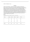
In image on the right clear differences are seen. Osteoporotic femur is less dense and even has gaps, a
different microarchitecture is seen. But at the highlighted region, it looks very similar to healthy bone.
This is the region that carries the most load. So bone mass is lost at regions that are less important, but
bone tries to remain density in important load bearing regions. So for normal loading conditions the bone
is not even that much less strong (but if you have odd loading conditions, like when you fall, then healthy
bone will be strong enough to withstand forces while osteoporotic bone will not).
So when looking at microarchitectural changes and loss of bone mass, it is
understandable that there is increased fragility. But this is not
quantifiable, so the second definition was made.
Bone mass
• Changes with age
• Peak bone mass at age of 30
• Males have higher peak than females
• From there on you will lose bone mass
4
Lecture 1 – Basics & clinical issues
BASICS
Different levels of organization
• Femur is biggest bone
• In center it consists of dense material that forms hollow tube, called cortical/compact bone
o Has some porosity, but very small (too small for naked eye)
• Near end it is more filled with porous type of bone, called
trabecular/cancellous/spongy bone
o Consists of struts of bone material called trabeculae
o Spacing ~1 mm
Trabecular bone
• Longitudinal organization in trabeculae is seen
• This is due to platelet like structures in trabeculae that are laid down by
cells in direction of trabeculae
• In tissue you have osteocytes located in small spaces called lacunae
within bone tissue.
• Osteocytes have a lot of process, they connect to other osteocytes.
o Processes are laid down in canals, called canaliculi.
Compact bone
• Cylindrical structures called osteons
• At center of osteon you have cavity, the central canal. Here you
have nerves and blood vessels.
• Osteocytes cells are present within an osteon, here they are in
their lacuna and connected via canaliculi (similar to what is seen
in trabecular bone).
Different length scales
• At meter, largest bones in body such as femur. So organ level
• At millimeters, you look at cancellous or cortical bone regions.
• At micrometers, at length scale where you recognize cells (~10 μm)
• At nanometers, actual tissue material in cancellous and cortical bone is more or less the same and
consists of collagen and hydroxyapatite.
Focus is on mechanics: why?
Mechanical loading plays an important role during bone
• Formation
• Growth
• Adaptation
• Functioning
1
,Formation & development
Formation
• At embryonal stage, bone is nothing more than cartilage plug
• Cartilage plug starts to mineralize at center at some point
• Mineralization extends plug gets bigger, at some point vascularization happens turns in real
bone tissue
• Now you have bone tissue with cartilage plugs at the
ends
• When bone further grows you get secondary
mineralization zone in cartilage ends that are left,
this gets penetrated by vessels and then turns into
real bone.
• In center you have bone, cartilage between bone
parts become growth plate and outer cartilage
layers turn into the joint surface.
Growth
• Growth plate consists of hypertrophic chondrocytes
• As hypertrophy of the chondrocytes occur, the bone on top is
being pushed away further outwards bone grows
• After hypertrophy, they mineralize the surroundings.
• Mineralized cartilage is turned into real bone
• Around age of 17 growth plate is closed and growth stops
Adaption
• Bones are not yet inert, they can still adapt.
• Trabecular architecture can adapt, e.g. if you load your bone more it becomes more dense.
• Can also be mis adaption, resulting in joint/bone diseases
During all these stages, mechanical factors play an important role.
Formation
• From stem cells tissue can develop
• Depending on strain and hydrostatic pressure different types
of tissue can develop
• Mainly hydrostatic pressure tissue becomes cartilage
• Mainly strain fibrous connective tissue
Development
• Bone structure of tibia in pig: trabeculae
o At 6 weeks disorganized
o Over time structure organizes itself
o Becomes more vertically oriented
• Human femur
o Trabeculae organization is not random
o Preferred orientation in certain direction at
different locations due to mechanical loading
2
,Remodeling & adaption
• Wolff’s law: mass and orientation of trabecular structure are adapted to its mechanical load.
• If a bone fractures it might grow back together and form new optimized architecture structure.
• Bone tissue is constantly renewing itself: bone turnover
• Micro-cracks are repaired
• Enables adaptation of the structure
Trabecular bone
• Osteoclasts reserve bone tissue, can dig cavity in bone tissue
• Osteoblasts: deposit bone tissue in this cavity
• By doing so you get new bone tissue
Cortical bone
• Osteoclasts dig tunnel in direction of longitudinal axis of bone
• Osteoblasts follow osteoclasts and fill tunnel again, but leave small
center that can be filled by blood vessels and nerves.
CLINICAL ISSUES
Skeletal deformities
• Can be due to misalignment of force
• Mechanical loading plays an important role during growth
• A growth plate normally aligns perpendicularly to the force that is
acting on the bone, so if force is on different angle, then growth plate is at different angle which
results in growth of bone in different direction.
• Aside from orientation, the magnitude of force is also important for bone development. If there
is very little force, then not a lot of bone development.
• Idiopathic deformities: even when orientation and magnitude is normal, you can get skeletal
deformities. An example is scoliotic spine, the vertebrae deform force not straight anymore.
Delayed fracture healing
Normally fracture heals very well
• For long bone, when fracture has taken place a blood clot is formed, called hematoma. If you
stabilize bone (e.g., with splint), the hematoma will transform into cartilage further stabilize
region then mineralize then bone repaired
• Bones can heal without scars.
But, sometimes fractures don’t heal
• Happens if there is too much or not enough motion
• Type of fixation has important effect
o Not too flexible
o Not too rigid
• Cyclic axial motion before initiation of ossification more rapid healing
• Strain rate matters for healing process
Implant loosening
• Joint replacement implants
• Limited life time of prostheses
3
, Hip implants
• Tries to stimulate bone in growth in the implant
• Major problems:
o Insufficient/unsuccessful ingrowth of bone loosening of
implant, so replacement is needed.
o Bone resorption around implant, due to stiffness of implant so
unloading of surrounding bone.
• Failure: determined by mechanical factors
Osteoarthrosis
• Disease of whole joint
• Cartilage is ‘visualized’ by joint space. If x-ray does not
show joint space, then cartilage is gone.
• Without cartilage, you get bone on bone interactions
Degeneration of cartilage is seen, but also changes to subchondral
bone and synovial fluid. Hence, whole joint disease.
OSTEOPOROSIS
Definitions
Two definitions:
1. Affection of the skeleton characterized by:
• Low bone mass
• Microarchitectural changes
• Increased fragility
Problem with this definition, not quantifiable.
2. WHO definition:
• Bone mineral density (BMD) less than 2.5 SD from the average at the age of 30.
With first definition, the main issue is increased fragility. Because low bone mass is not necessarily a
problem. That your microarchitectural looks less nice is not big issue either as you don’t see it anyways.
But increased fragility is the problem with osteoporosis.
In image on the right clear differences are seen. Osteoporotic femur is less dense and even has gaps, a
different microarchitecture is seen. But at the highlighted region, it looks very similar to healthy bone.
This is the region that carries the most load. So bone mass is lost at regions that are less important, but
bone tries to remain density in important load bearing regions. So for normal loading conditions the bone
is not even that much less strong (but if you have odd loading conditions, like when you fall, then healthy
bone will be strong enough to withstand forces while osteoporotic bone will not).
So when looking at microarchitectural changes and loss of bone mass, it is
understandable that there is increased fragility. But this is not
quantifiable, so the second definition was made.
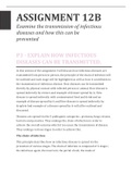
Bone mass
• Changes with age
• Peak bone mass at age of 30
• Males have higher peak than females
• From there on you will lose bone mass
4