100% satisfaction guarantee Immediately available after payment Both online and in PDF No strings attached
logo-home
Samenvatting Biogenie 5.2 Hoofdstuk 1 - functionele morfologie van de cel $6.89
Add to cart

Summary

Samenvatting Biogenie 5.2 Hoofdstuk 1 - functionele morfologie van de cel

1 review
 139 views  2 purchases
  • Course
  • Institution
  • Book

Volledig thema 1 is samengevat.

Preview 2 out of 7  pages

  • No
  • Hoofdstuk 1
  • March 19, 2021
  • 7
  • 2020/2021
  • Summary
  • Secondary school
  • KSO
  • 5

1  review

review-writer-avatar

By: karindiels • 2 year ago

Translated by Google

too few

avatar-seller
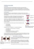
Thema 1: functionele morfologie van de cel


1. Cellen in relatie met andere organisatieniveaus
Biosfeer: gedeelte van de aarde waar leven mogelijk is. Georganiseerd in verschillende ecosystemen
waarin populaties van allerlei soorten organismen voorkomen.
Organisatieniveaus:

Supramacroscopische Biosfeer → ecosysteem → populatie
organisatieniveaus
Macroscopische organisatieniveaus Organisme → orgaanstelsel → orgaan → weefsel
(blote oog – loep)
Microscopische organisatieniveaus →cel
(lichtmicroscoop)
Submicroscopische →celorganel → macromolecule
organisatieiveaus
(elektronenmicroscoop)
Moleculaire organisatieniveaus →molecule → atoom
(indirecte methodes –
kristallografie)
2. Observeren van cellen

 Vanaf 17de eeuw: loepen
 Vanaf 19de eeuw: Organismen bestaan uit cellen (Schleiden en Schwann)
 Vanaf 20ste eeuw: verfijnd onderzoek (lichtmicroscoop en elektronenmicroscoop)
❖ Met BLOTE OOG
Eicel kikker eicel vogel


❖ Met LICHTMICROSCOOP

 Celstructuren waarnemen (celwand, celmembraan, cytoplasma, vacuole, zetmeel-en bladgroenkorrels)
 Planten- en dierencellen 10 à 200 µm
Epidermis uirok bloedcellen


❖ Met ELEKTRONENMICROSCOOP

 Sinds 1933
 Submicroscopisch: zo klein dat je ze met een lichtmicroscoop niet kan waarnemen
 1 nanometer
 submicroscopische structuur zien: celorganellen

, Oplossend vermogen (optische instrumenten)

 = scheidend vermogen, resolutie
 drukt uit Hoe dicht punten bij elkaar kunnen liggen om nog net afzonderlijk te zien
 Het oog: 0,2 mm
 Lichtmicroscoop: afstand tussen 2 punten niet kleiner dan de halve golflengte van het gebruikte licht
 Elektronenmicroscoop: 0,2 nm & altijd in grijstinten , 2 soorten:
Transmissie-elektronenmicroscoop (TEM): doorsnede
rasterelektronenmicroscoop (REM): oppervlak

3. Submicroscopische studie van het celoppervlak

Celwand (enkel bij planten, fungi en bacteriën)
Bouw:
✓ wand rond de plantencel, gebouwd uit vezeltjes (cellulose: vezelige moleculen in lagen en vormen
netwerken met mazen ertussen)
✓ kan zeer dik zijn (hout)
✓ Fungi: chitine
✓ Bacteriën: mucopeptide (‘achillespees’ van de bacterie) (penicilline kan mucopeptide verstoren)
✓ Glycocalyx: slijmerige laag bovenop bacterien celwand
Functie:
✓ Stevigheid van de plantencel
✓ Geeft vaste vorm aan de plantencel
✓ Beschermende laag (afbraak, ongunstige omstandigheden)

Celmembraan/ plasmalemma
 Cytoplasma: geleiachtige vloeistof (cytosol) + celstructuren
 Cytosol: water, eiwitten, suikers, vetten, nucleïnezuren, ionen.
Bouw:
✓ Lipidendubbellaag opgebouwd uit fosfolipiden, cholesterol en proteïnen daartussen (perifere (op
dl) en transmembraanproteïnen (door dl))
✓ Extracellulaire (buiten) kant: sacharideketens op lipiden en proteïnen = glycolipiden en
ki,i,iglycoproteïnen
✓ Fosfolipidenmoleculen: gevorkte staart is apolair en hydrofoob (waterafstotend) en kop is polair en
zzzzhydrofiel → dubbellagen, staarten naar elkaar


Fosfolipidendubbellaag: een rangschikking van moleculen die vrij kunnen bewegen in het vlak van de
dubbellaag.

The benefits of buying summaries with Stuvia:

Guaranteed quality through customer reviews

Guaranteed quality through customer reviews

Stuvia customers have reviewed more than 700,000 summaries. This how you know that you are buying the best documents.

Quick and easy check-out

Quick and easy check-out

You can quickly pay through credit card or Stuvia-credit for the summaries. There is no membership needed.

Focus on what matters

Focus on what matters

Your fellow students write the study notes themselves, which is why the documents are always reliable and up-to-date. This ensures you quickly get to the core!

Frequently asked questions

What do I get when I buy this document?

You get a PDF, available immediately after your purchase. The purchased document is accessible anytime, anywhere and indefinitely through your profile.

Satisfaction guarantee: how does it work?

Our satisfaction guarantee ensures that you always find a study document that suits you well. You fill out a form, and our customer service team takes care of the rest.

Who am I buying these notes from?

Stuvia is a marketplace, so you are not buying this document from us, but from seller denissteffie. Stuvia facilitates payment to the seller.

Will I be stuck with a subscription?

No, you only buy these notes for $6.89. You're not tied to anything after your purchase.

Can Stuvia be trusted?

4.6 stars on Google & Trustpilot (+1000 reviews)

47561 documents were sold in the last 30 days

Founded in 2010, the go-to place to buy study notes for 15 years now
Start selling
$6.89  2x  sold
  • (1)
Add to cart
Added