Chapter 15- OncologyMed Surg Study Guide Exam 1
Warning Signs of Cancer- CAUTION
C- changes in bowel or bladder A- sore that does not heal
U-unusual bleeding or discharge
T- thickening or a lump
I-indigestion or difficulty swallowing O- obvious change in a wart or mole N- nagging cough or hoarseness
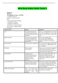
Characteristics Benign Malignant
Cell Well- differentiated cells resemble normal cells of the
tissue from which the tumor
originatedCells are undifferentiated and may
bear little resemblance to the normal
cells of the tissue from which they
arose
Mode of Growth Tumor grows by expansion and does not infiltrate the surrounding tissues, usually
encapsulatedGrows at the periphery and overcomes contact inhibition to invade and infiltrate surrounding tissues
Rate of Growth Rate of growth is usually slowRate of growth is variable and depends
on the level of differentiation, the
more anaplastic
Metastasis Does not spread by metastasisGains access to the blood and lymphatic
channels and metastasizes to other areas of the body or grows across body cavities such as the peritoneum
General Effects Usually localized phenomenon that does not cause generalized effects unless its location interferes with vital functionsOften causes generalized effects such as anemia, weakness, systemic inflammation, weight loss, and CACS (cancer-related anorexia- cachexia syndrome)
Tissue Destruction Does not usually cause tissue
damage unless its location interferes with blood flowOften causes extensive tissue damage as the tumor outgrows its blood supply or encroaches on blood flow to the area; may also produce substances that cause cell damage
Ability to Cause Death Does not usually cause
death unless its location
interferes with vital
functionsEventually causes death unless growth can be controlled Causes
●Viruses and Bacteria
○HPV (Human Papillomavirus Infection- dirty sex and leaves warts) cervical cancer, Hepatitis B or C Liver cancer, H. Pylori GI/colorectal cancer
●Physical Agents
○Sunlight, radiation, chronic irritation/inflammation
○Fair-haired/skin, blue/green eyes, occupation (outside etc.), tanning bed use, Radiation from CXR increased risk of lung cancer
●Chemical Agents
○Tobacco smoke, industrial chemicals and asbestos (lung cancer)
●Genetics and Familial Factors
○Hereditary CA syndrome - Colorectal, breast, ovarian, prostate
○Shared environments/lifestyle
●Dietary Factors
○Fats, alcohols, salt-cured, smoked, red/processed meats
○Protective foods- fruits, veggies, whole grains
●Hormonal Agents
○DES (Diethylstilbestrol- estrogen medication used to treat some cancers and maintain pregnancy but led to breast and ovarian cancer later, DES babies carry increased risk of cancer), hormonal changes, hormone replacement therapy
●Chance
○Some families and some not
●Health Promotion
○Healthy weight, physically active lifestyle, eat plenty of fruits, veggies, and whole grains
Detection and Prevention
●Primary
○Health promotion and risk reduction strategies
○Before you even have cancer, stay away from carcinogens, take Tamoxifen to decrease chances of breast cancer
●Secondary
○Screening and early detection activities, prostate exam for men
●ACS (American Cancer Society) Screening Guidelines
○Breast- mammography, Cervix- Pap test every year, HPV DNA test, Colorectal- fecal occult blood test, colonoscopy, may remove polyps as protection, Endometrial- women at menopause should report bleeding or spotting, Lung- Low-dose CT, Prostate- 50+, digital rectal exam (DRE) and prostate-specific antigen (PSA)
●Tertiary
○Monitor for and preventing recurrence, post cancer have high recurrence risk
Diagnostics
●Tumor marker identification
○If there is a tumor increase tumor marker, CA 125 shows increased risk of cancer
●Mammography
●MRI, CT scan, Fluoroscopy (real time moving x-ray movie), Ultrasound
○CT scan contrast allergy, shellfish/iodine allergy, kidney function, MRI- no metal, assess claustrophobic patient
●Endoscopy ○In with scope, lung, bleeding, tubing, GI both upper and lower
●Nuclear Imaging, PET Scan
○Radiographic isotopes can detect where cancer is at, travels to hot spot areas (red color on image), or infection, helps find primary source
●Tumor Staging and Grading
○TNM (T= extent of primary tumor, N= the absence or presence and extent of regional lymph node metastasis, M= absence or presence of distant metastasis
■Tx= tumor cannot be assessed, T0= no evidence of primary tumor, Tis= carcinoma in situ ?????????? T1-4= increasing size and/or local extent of the primary tumor
■Nx= regional lymph nodes cannot be assessed, N0= no regional lymph node metastasis, N1-3= increasing involvement of regional lymph nodes
■Mx= distant metastasis cannot be assessed, M0= no distant metastasis, M1= distant metastasis
Biopsy
●Excisional
○Small, easily accessible tumors of the skin, upper/lower GI and upper respiratory tract, done outpatient, biopsy takes time to read
●Incisional
○Larger mass, wedge of tumor removed, colon, breast lumpectomy
●Needle
○Easily and safely accessible masses, breast, thyroid, lung, liver, kidney
○Outpatient basis, local anesthetic used, monitor site for infection
Treatment (Malignant)
●Surgery
○Remove entire tumor or as much as possible (debulking) as well as surrounding tissue including regional lymph nodes, clear margins, clear out any cancer cells
○Local vs Wide excision
○Minimally invasive techniques= decreased trauma, blood loss, risk for infection, complications, surgery time, anesthesia, and increased recovery time
○Video-assisted endoscopy and laparoscopic robotic-assisted
●Prophylactic
○Colectomy, mastectomy, oophorectomy (removal of ovaries), preventative, remove before cancer develops, polyps, inflammatory bowel disease can have portion of bowel removed to decrease risk of cancer
●Palliative
○Relieve symptoms, provide comfort, promote quality of life, not helpful in the treatment
○DNR code, labs and diagnostics- blood sugar, vital signs- PRN, activity- as tolerated/what patient wants, diet- what they can tolerate, accommodate, homemade foods, medications- pain med, morphine, NV, scopolamine patch
●Reconstructive
○Breast, head, neck, and skin cancer, increase self esteem, reconstruction is done during surgery and post-op
○
●Nursing management ○Kind, caring, involve family, monitor risk for infection
Radiation
●Teletherapy(external beam)
○Most common
○Daily fractions accurately target the tumor
○Typically 5 days a week M-F for 4-6 weeks
●Brachytherapy(internal)
○High, intense dose
○Dose delivered to localized area-via needles, catheters and other methods
○Affects the skin- very radioactive, GI tract, bone marrow-causes anemia
●Toxicity
○Alopecia, stomatitis, A/N/V/D, fatigue, thrombocytopenia, radiation dermatitis, anemia
Nursing management
●Precautions
○BR, private room
○IDC, low residue diet-slows stool to keep from having a BM
○Distance-6ft
○Shielding
○Time-30 mins/day
●Protecting caregivers
○Wear dosimeter badge to detect radiation exposure
○No pregnant staff/visitors
○No children visitors
Chemotherapy- use of antineoplastics in attempt to destroy cancer cells.
●Systemic
○IV, IM, PO, Topical, Intracavitary
○Chemo is given in “rounds”
○20-90% of the tumor cells are destroyed-each time a tumor is exposed to chemo
●Extravasation
○Vesicant agents cause tissue damage/possible necrosis
●Toxicity
○GI, hematopoietic, renal, cardiopulmonary, fatigue, reproductive, cognitive- “chemo fog” or “chemo brain”
Nursing management
●Assess fluid/lytes
●Assess cognitive status- no falls, safety
●Decrease risk for infection & bleeding- avoid large crowds, wear mask, use soft bristle toothbrush
●Administering chemo- double glove, gown, - meds need to be double bagged, spill kit for floor
●Manage fatigue- body mechanics, evaluate energy levels, weekly planning, attentiveness to personal situation
●Rest and exercise need to be prioritized
●Prevent N/V
Hematopoietic stem cell transplant


