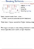
3 layers to the heart Conduction of cardiac action Heart innervated by both:
- Epicardium (outer) potentials - the parasympathetic NS –
- Myocardium Cardiomyocytes have intercalated Ach (vagus nerve X
- Endocardium disks between them: innervation of SA and AV
Pericardium surrounds the heart – - Interconnect cardiac muscle nodes) decreases
made of parietal and visceral cells contractility
(epicardium). Cavity contains - Secured by desmosomes - the sympathetic NS –
pericardial fluid -lubrication - Linked by gap junctions NA/A β1 adrenergic
Too much fluid = pericarditis o Propagate action innervation of SA and AV
potentials in nodes and ventricular
adjacent cells. muscle increases
o Local changes in contractility.
currents -passive
depolarisation of Cardiac output = HR x SV
adjacent muscle Stroke volume = EDV - ESV
cells (voltage gated Preload = EDV
ion channels) Increases in EDV lead to increases
through gap in contractility and more venous
junctions return (so higher sarcomere
Action potentials are propagated length -> more force before
Left ventricle has a higher pressure along the sarcolemma -> Na+ influx - contraction) -> increases SV ->
(4-6 times)than the right (3:1 ratio in > can go through gap junctions -> increases CO.
muscle mass) due to it having a depolarise next cell Changes due to:
thicker wall. Peristaltic motion - Physical factors – more
BP = systolic/diastolic pressure Helps to force blood into the right optimum myofilament
Excitation-contraction coupling direction. (ventricles contract from overlapping -> decrease
Cardiomyocytes create an action the apex up). 80% of blood naturally lattice and myofilament
potential in the SA node -> only goes into the next cavity of the spacing -> increased
pathway though is via AV node -> heart due to pressure changes, but probability of interaction
bundle of His -> Purkinje fibres -> the peristaltic motion helps to pump between contractile
cardiomyocytes. out the last 20% of CO. (but note components
In cardiomyocytes: that the ventricles don’t completely - Activating factors – increase in
AP at T-tubule causes Ca2+ influx empty) Ca2+ sensitivity and Ca2+
from L-type Ca2+ voltage-gated release
channels and Ca2+ binds to the Contractility
ryanodine receptors (RyRs) -> Three factors affect ESV:
Affected by sympathetic drive to
calcium induced calcium release -> ventricular muscle fibres (NA at β1) Preload : higher EDV increases
Ca2+ binds to troponin in sarcomere And hormonal control (circulating A contractility
unit -> myosin & actin cross bridges. and NA. Contractility : increases SV
Quantification of contractility = Afterload : pressure in which the
Anything that alters Ca2+ release or Ejection fraction = ratio of stroke heart has to pump against. Higher
storage alters volume to end diastolic volume pressure in the aorta = more
contractility/relaxation. E.g. Ca2+ EF = SV/EDF (percentage of blood work/force required by the heart.
channel blockers, β blockers, pumped out per beat. <40% = HF Afterload increases = CO
caffeine. decreases unless the heart puts
more work in.
Heart rate control:
Neuronal end endocrine
- Sympathetic = positive chronotropic factors (NA & A)
- Parasympathetic = negative chronotropic factors (Ach – muscarinic)
Atrial reflex/Bainbridge reflex
Adjusts HR in response to venous return
- Sympathetic makes it easier to reach threshold – reduces repolarisation
- Parasympathetic makes it harder to reach the threshold – decreases
membrane potential – more time needed to reach threshold.
- (NA & A binding increases renin)