Recombinant DNA
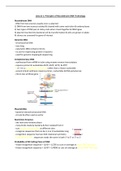
- DNA from two sources usually one is a plasmid
1) DNA from two sources isolated & cleaved with same restriction & endonuclease
2) two types of DNA pair at sticky ends when mixed together & DNA ligase
3) plasmid inserted into bacterial cell by transformation & cells are grown on plate
4) clones are screened for gene of interest
Genomic DNA
- chromosomal DNA
- very long
- eukaryotic DNA contains introns
- no use for expressing protein in bacteria
- used for genome mapping & sequencing
Complementary DNA
- synthesised from mRNA in tube using enzyme reverse transcriptase
- requires primer & nucleotides dATP, dGTP, dCTP, & dTTP
d = it is a deoxyribose nucleotide rather than a ribose nucleotide
- second strand synthesis requires primer, nucleotides & DNA polymerase
- intron-less artificial gene
Plasmid DNA
- bacterial extrachromosomal DNA
- circular & often used as vector
Restriction Enzymes
- aka restriction endonuclease
- many kinds made by bacteria & then isolated from it
- cuts double stranded DNA at different sites
- recognise sequences that can be 4, 6 or 8 nucleotides long
- recognition sequence has two-fold rotational symmetry
palindromic sites: sequences reads the same in both 3’ to 5’ or 5’ to 3’
Probability of RE Cutting Piece of DNA
- 4 base recognition sequence = (1/4)4 = 1/256 so cuts on average at every 256th base pair
- 6 base recognition sequence = (1/4)6 = 1/4096 so cuts on average at every 4096th base pair
,Blunt & Sticky Ends
- blunt ends are when DNA is cut evenly
not used often
- sticky ends are result of DNA not cutting cleanly resulting in exposed bases
using DNA ligase on sticky ends allows them to reform circular DNA
used often as it easier to slip DNA of interest onto sticky ends
Restriction Enzymes Cut Sugar Phosphate Backbone
- cut the sugar backbone leaving 5’ phosphate & 3’ OH
- remaining segments used by DNA ligase to re-join DNA
Joining DNA
- requires DNA ligase that ligates the phosphodiester backbone
ligate: joining molecules together with new chemical bond
- ligase joins 5’ phosphate to 3’ OH in double stranded DNA
Vectors in Cloning Experiments
- DNA fragments joined to vector
- relatively small DNA molecule usually with single site for chosen restriction enzyme
- capable of replicating in host organism
- contains desirable gene e.g. antibiotic resistance
Hosts
- bacteria, yeast, animal cells in culture etc
- can be easily grown in large quantities & must be non-pathogenic
- only fraction of host cells take up plasmid during transformation
- not all plasmids contain insert – a re-ligation of vector
can be minimised by treating linearized vector with phosphatase
Blue-White Screening
- allows identification of it antibiotic resistant cells contain recombinant DNA
- vector contains genes coding for enzyme β-galactosidase (lacZ)
- this enzyme converts substrate X-gal into blue coloured product
- if gene coding for the enzyme is disrupted, no enzyme would be produced and colony would be
white
blue product = is recombinant clone
no blue product = not recombinant clone
Library
- large no. of different genomic fragments of cDNAs inserted into vector
- mixture of recombinant DNAs
,- each colony is derived from single recombinant DNA molecule
- library becomes collection of clones
, Lecture 2: Gene Analysis
Agarose Gel Electrophoresis
- common technique used to separate DNA fragments by size
- uses gel made from polymer e.g. polysaccharide agarose
- agarose gel is 3D matrix of interconnected agarose molecules that form channels & pores that
biomolecules pass through
pore sizes are determined by conc of agarose gel
- gel acts as molecular sieve to separate nucleic acids on basis of size by slowing movement of longer
molecules so they separate from shorter ones
- electric charge is applied and fragments are separated by electrochemical properties
- nucleic acids carry negative charges on phosphate groups and so will travel to positive pole in
electric field
- tube contains mixture of DNA molecules with different sizes, a dye that lets us see tube contents
and glucose & sucrose to make solution load into wells
- fluorescent dye e.g. ethidium bromide is used as it’s visible under UV
- fluorescent molecule intercalates into double helix and allows us to see band patterns
corresponding to the DNA
- molecular weight marker which is piece of DNA cut with restriction enzymes & gives fragments of
known size is added so we can compare size of DNA we’re measuring in electrophoresis
- a buffer is used to allow the current to pass through the gel and prevent overheating & melting of
the agarose gel
correct amount of buffer required to allow good migration of DNA
- to load DNA sample into wells, a loading buffer containing some sugar & glycerol is added to make
loading easier & prevent DNA diffusing into buffer and ensuring it remains in well
Restriction Mapping
- restriction fragment analysis can rapidly provide info of DNA sequence
- if we have DNA fragment of 10kbs that’s cut with two different restriction endonucleases, enzyme
A will cut DNA & generate two fragment of 2kbs & 8kbs whilst enzyme B recognises sequence in
DNA and cuts once at 5kbs generating fragments of same size
- if we combine both enzymes it will generate three observable fragments; 2kbs, 3kbs & 5kbs
- if we run DNA fragments in gel for enzyme, we would detect it by running it on gel corresponding
to fragment of 2kbs & 8kbs
- for enzyme B, it’ll generate two fragments at equal sites and will only be detected in gel as single
bond as they’re same size but band will contain two fragments at 5kbs
Restriction Fragment Analysis
- used to compare two different DNA molecules e.g. two different alleles for a gene
- if nucleotide difference affects recognition site of restriction enzyme, a change in one nucleotide in
restriction enzyme could prevent it from cutting DNA
- polymorphisms: variations in DNA sequences
- polymorphisms impact recognition site of restriction enzymes and are called restriction fragment
length polymorphisms
- you can use RFLP analysis to determine differences in alleles
- for example, mutation in β-globin gene leads to defective protein causing sickle cell disease
- you can use RFLP to differentiate between normal allele & mutant allele that causes defective
protein in SCA


