100% satisfaction guarantee Immediately available after payment Both online and in PDF No strings attached
logo-home
Samenvatting Circulatie Kind en Neonaat 129 - IC Neonatologie $6.96   Add to cart

Summary

Samenvatting Circulatie Kind en Neonaat 129 - IC Neonatologie

 50 views  2 purchases
  • Course
  • Institution
  • Book

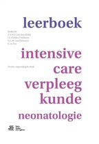
Deze samenvatting is gemaakt ter voorbereiding voor het 'Circulatie 129 - Kind en Neonaat' tentamen voor de opleiding Intensive Care Neonatologie (ook bruikbaar voor de opleiding High Care Neonatologie en Intensive Care Kinderen) in mei 2021 vanuit de Erasmus MC Academie. De theorie is afkomsti...

[Show more]
Preview 3 out of 28  pages

  • Yes
  • November 11, 2021
  • 28
  • 2021/2022
  • Summary
avatar-seller
Circulatie 129 – Kind en neonaat
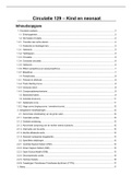
Inhoudsopgave
1. Circulatoir systeem......................................................................................................................................3
1.1. Embryogenese.......................................................................................................................................3
1.2. De foetale circulatie...............................................................................................................................4
1.2.1. Transitie naar extra-uterien....................................................................................................................4
1.3. Anatomie en fysiologie hart...................................................................................................................5
1.3.1. Hartwand................................................................................................................................................5
1.3.2. Hartkleppen............................................................................................................................................5
1.3.3. Circulatie................................................................................................................................................5
1.3.4. Coronaire circulatie................................................................................................................................6
1.3.5. Harttonen...............................................................................................................................................6
1.3.6. Effect sympathicus en parasympathicus...............................................................................................7
1.3.7. Bloeddruk...............................................................................................................................................7
1.4. Pompfunctie...........................................................................................................................................8
1.4.1. Preload en afterload...............................................................................................................................8
1.4.2. Frank Starling Curve..............................................................................................................................9
1.4.3. Veneuze return.......................................................................................................................................9
1.4.4. Compensatiemechanisme.....................................................................................................................9
1.5. Basisobservaties....................................................................................................................................9
1.6. Arteriële drukcurve...............................................................................................................................11
1.6.1. Gedempte curve...................................................................................................................................11
1.6.2. Hoge curve (ringing-curve / overshoot-curve).....................................................................................12
2. Aangeboren hartafwijkingen......................................................................................................................12
2.1. Abnormale ontwikkeling van de grote vaten.......................................................................................12
2.1.1. Coarctatio aortea.................................................................................................................................12
2.1.2. Dubbele aortaboog..............................................................................................................................12
2.1.3. Abnormale oorsprong van de rechter arteria subclavia......................................................................12
2.1.4. Interruptie van de aorta........................................................................................................................13
2.1.5. Rechts afdalende aorta........................................................................................................................13
2.1.6. Aberrant verlopende longarteriën........................................................................................................13
2.2. Specifieke afwijkingen.........................................................................................................................13
2.2.1. Ventrikel Septum Defect (VSD)...........................................................................................................13
2.2.2. Atrium Septum Defect (ASD)...............................................................................................................13
2.2.3. Open Ductus Botalli (ODB)..................................................................................................................13
2.2.4. Decompensatio cordis.........................................................................................................................15
2.2.5. Hydrops foetalis...................................................................................................................................16
2.2.6. Tweelingen-Transfuseur-Transfusée-Syndroom (TTTS)....................................................................17
3. Ritme..........................................................................................................................................................17

, 3.1. Actiepotentiaal.....................................................................................................................................17
3.2. Geleidingssysteem..............................................................................................................................18
3.3. Driehoek van Einthoven.......................................................................................................................18
3.4. Weergave van de geleiding op het ECG.............................................................................................19
3.5. Normaalwaarden..................................................................................................................................19
4. Hartritmestoornissen.................................................................................................................................19
4.1. Sinusaritmieën.....................................................................................................................................20
4.1.1. Sinusbradycardie.................................................................................................................................20
4.1.2. Sinustachycardie..................................................................................................................................20
4.1.3. Sinusaritmie.........................................................................................................................................20
4.1.4. Sinusarrest...........................................................................................................................................20
4.2. Supraventriculaire ritme- en geleidingsstoornissen............................................................................20
4.2.1. Atriumflutter..........................................................................................................................................20
4.2.2. Atriumfibrilleren....................................................................................................................................20
4.2.3. Supraventriculaire tachycardie (SVT)..................................................................................................21
4.3. Atrioventriculaire geleidingsstoornissen..............................................................................................21
4.3.1. Eerstegraads AV-blok..........................................................................................................................21
4.3.2. Tweedegraads AV-blok........................................................................................................................21
4.3.3. Derdegraads AV-blok (totaal AV-blok)................................................................................................21
4.3.4. AV-junctional ritmes.............................................................................................................................21
4.4. Ventriculaire ritme- en geleidingsstoornissen.....................................................................................21
4.4.1. Prematuur atriaal complex (PAC)........................................................................................................21
4.4.2. Prematuur ventriculair complex (PVC)................................................................................................21
4.4.3. Prematuur junction complex (PJC)......................................................................................................21
4.4.4. Ventriculaire tachycardie......................................................................................................................22
4.4.5. Ventrikelfibrilleren................................................................................................................................22
5. Shock.........................................................................................................................................................22
5.1. Hypovolemische shock........................................................................................................................23
6.4. Obstructieve shock..............................................................................................................................23
6.5. Cardiogene shock................................................................................................................................23
6.6. Distributieve shock...............................................................................................................................24
6. Medicamenten...........................................................................................................................................24
6.1. Werking inotropica en chronotropica...................................................................................................24
7. Elektrolyten en vocht.................................................................................................................................25
7.1. Elektrolyten..........................................................................................................................................26
7.2. Kenmerken verschillende elektrolytenstoornissen..............................................................................26
7.2.1. Natrium.................................................................................................................................................26
7.2.2. Kalium..................................................................................................................................................27
7.2.3. Calcium................................................................................................................................................27
7.2.4. Fosfaat.................................................................................................................................................27
7.2.5. Magnesium...........................................................................................................................................27


2

, 7.2.6. Ureum...................................................................................................................................................27
7.2.7. Creatinine.............................................................................................................................................28
7.3. Relatie elektrolytstoornis en kenmerk op het ECG.............................................................................28
7.4. Verpleegkundige bewaking en interventies.........................................................................................28


1. Circulatoir systeem
1.1. Embryogenese
Tijdens de ontwikkeling van een embryo volgen een aantal fasen zich op: morula, blastula en granulatie.
Tijdens de granulatie wordt een drielagig kiemblad gevormd bestaande uit endoderm, mesoderm en
ectoderm. De hartspiercellen worden gevormd vanuit het mesoderm. Het hart begint zich te vormen rond
de 16e dag en bestaat dan uit een buis, de primaire hartbuis met waarneembare spiercontracties. Het
primitieve hart begint op dag 24 te kloppen en foetale harttonen zijn bij 10-12 weken hoorbaar. De
primaire hartbuis maakt een draaiing waardoor het in een lus komt te liggen. Tijdens het uitgroeien van
deze lus ontstaan een primitief atrium en ventrikel. De uitgang van het ventrikel heet truncus
arteriosus, deze uitgang splitst zich in de aorta en de a. pulmonalis (longslagader). De aorta splitst
zich nu in meerdere takken, kiewboogarteriën.

De primaire hartbuis is omgeven door een endotheelwand met daartussen een geleiachtige substantie
wat zich vult met mesenchymcellen, hieruit worden de 3 lagen van het hart gevormd: endocard –
myocard – pericard. Er ontstaan verdikkingen vanuit de achter- en voorwand van het endocard
(endocardkussens) welke de grens tussen het atrium en ventrikel vormt. Deze endocardkussens groeien
samen en ontwikkelen zo het linker en rechter atrioventriculaire kanaal. In dezelfde periode (vanaf de
5e week) groeit vanuit het atrium een septum (tussenschot) in de richting van de endocardkussens. Dit
zorgt voor een scheiding tussen het linker- en rechteratrium. Het septum ontwikkelt zich zodanig dat het
bloed wel van rechts naar links, maar niet van links naar rechts kan stromen. Voordat de scheiding
compleet is, ontstaat er een gat in het septum, het foramen ovale. Later ontstaat er aan de linkerkant een
2e septum wat het foramen ovale sluit. Verstoringen in deze periode kunnen leiden tot diverse
septumdefecten.

De aorta splitst zich in meerdere takken (kiewboogarteriën). In het ventrikel gaat de scheiding tussen het
linker- en rechterharthelft anders. Het ventrikel groeit uit waarbij de onderkant van het hart niet van
afstand veranderd ten opzichte van de truncus arteriosus. Hierin moet ook een septum komen omdat er
nu 2 ventrikels zijn met 1 uitstroomkanaal. Dit septum groeit vanuit de a. pulmonalis en moet al
groeiende een draai maken om goede aansluiting op de ventrikels te verkrijgen. Daarom draait de aorta
om de arteria pulmonalis heen. Verstoringen in deze periode kunnen leiden tot een transpositie van de
grote vaten.

De kiewboogarteriën ontwikkelen
zich in de arcus aorta (aortaboog)
en de a. carotis (halsslagader). Er
is nog een 3e kiewboogarterie die zich
bevindt tussen de aorta en de arteria
pulmonalis, dit is de ductus
arteriosus (ductus van botalli). Het
hormoon prostaglandine houdt de
verbinding open voor de geboorte.

Bij 6 tot 8 weken heeft hart zijn
uiteindelijke vorm en zijn de kleppen
aanwezig.


3

The benefits of buying summaries with Stuvia:

Guaranteed quality through customer reviews

Guaranteed quality through customer reviews

Stuvia customers have reviewed more than 700,000 summaries. This how you know that you are buying the best documents.

Quick and easy check-out

Quick and easy check-out

You can quickly pay through credit card or Stuvia-credit for the summaries. There is no membership needed.

Focus on what matters

Focus on what matters

Your fellow students write the study notes themselves, which is why the documents are always reliable and up-to-date. This ensures you quickly get to the core!

Frequently asked questions

What do I get when I buy this document?

You get a PDF, available immediately after your purchase. The purchased document is accessible anytime, anywhere and indefinitely through your profile.

Satisfaction guarantee: how does it work?

Our satisfaction guarantee ensures that you always find a study document that suits you well. You fill out a form, and our customer service team takes care of the rest.

Who am I buying these notes from?

Stuvia is a marketplace, so you are not buying this document from us, but from seller larissarv. Stuvia facilitates payment to the seller.

Will I be stuck with a subscription?

No, you only buy these notes for $6.96. You're not tied to anything after your purchase.

Can Stuvia be trusted?

4.6 stars on Google & Trustpilot (+1000 reviews)

72042 documents were sold in the last 30 days

Founded in 2010, the go-to place to buy study notes for 14 years now

Start selling
$6.96  2x  sold
  • (0)
  Add to cart