NR 509 Week 3: The Musculoskeletal System
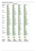
NR 509 Week 3: The Musculoskeletal System Table of Contents Note For learning purposes in this course, the weekly lessons are divided by body systems and focused on specific objective physical examination techniques. The lessons are adjunct to the assigned reading and by no means a substitution. Therefore, it is incumbent upon you to fully comprehend the associated subjective health history, health promotion, and anatomy and physiology as outlined in each textbook chapter. By the end of the course, you will be proficient with conducting a complete physical examination. Advanced Practice Assessment Techniques Assessment of the musculoskeletal system involves evaluating for issues of the bones, joints, skeletal muscles, ligaments and tendons, and cartilage throughout the body. As a clinician, you need to be aware of the expected anatomy and physiology of each structure within the musculoskeletal system, including the expected range of motion (ROM) of each joint. When abnormalities or limitations are discovered on assessment, diagnostic reasoning skills must be applied to determine how to best utilize the data. Your examination should be systematic. Include inspection, palpation of bony structures and related joint and soft tissue structures, assessment of range of motion, and special maneuvers to test specific movements. Recall that the anatomical shape of each joint determines its range of motion. There are two phases to range of motion: active (by the patient) and passive (by the examiner). There are specific differences in approach to assessment, as well as expected findings, across the lifespan. As you review the required readings this week, be alert for special considerations that relate to special patient populations. Review the videos below which demonstrate examination of the entire musculoskeletal systems and then practice your assessment skills. Continue to practice your skills until you can comfortably repeat a return demonstration of the entire assessment. The Hip Examination The hip joint is deeply embedded in the pelvis and is notable for its strength, stability, and wide range of motion. The stability of the hip joint, essential for weight bearing, arises from the deep fit of the head of the femur into the acetabulum, its strong fibrous articular capsule, and the powerful muscles crossing the joint and inserting below the femoral head, providing leverage for movement of the femur. Often, the examiner must assist the patient with movements of the hip, so further detail is provided below for flexion, abduction, adduction, and external and internal rotation. Meta-analyses suggest that no single test discriminates specific hip pathology. • Flexion: With the patient supine, place your hand under the patient's lumbar spine. Ask the patient to bend each knee in turn up to the chest and pull it firmly against the abdomen. Note that the hip can flex further when the knee is flexed because the hamstrings are relaxed. When the back touches your hand, indicating normal flattening of the lumbar lordosis, further flexion must arise from the hip joint itself. • Extension: With the patient lying face down, extend the thigh toward you in a posterior direction. Alternatively, carefully position the supine patient near the edge of the table and extend the leg posteriorly. • Abduction: Stabilize the pelvis by pressing down on the opposite anterior - superior iliac spine with one hand. With the other hand, grasp the ankle and abduct the extended leg until you feel the iliac spine move. This movement marks the limit of hip abduction. • Adduction: With the patient supine, stabilize the pelvis, hold one ankle, and move the leg medially across the body and over the opposite extremity. • External and internal rotation: Flex the leg to 90° at hip and knee, stabilize the thigh with one hand, grasp the ankle with the other, and swing the lower leg medially for external rotation at the hip, and laterally for internal rotation. Although confusing at first, it is the motion of the head of the femur in the acetabulum that identifies these movements. • The Knee Examination • The knee joint is the largest joint in the body. It is a hinge joint involving three bones: the femur, the tibia, and the patella (or knee cap), with three articular surfaces, two between the femur and the tibia and one between the femur and the patella. Note how the two rounded condyles of the femur rest on the relatively flat tibial plateau. There is no inherent stability in the knee joint itself, making it dependent on four ligaments to hold its articulating femur and tibia in place. This feature, in addition to the lever action of the femur on the tibia and the lack of padding from overlying fat or muscle, makes the knee highly vulnerable to injury. Learn to examine "the seven structures of the knee": the medial and lateral menisci, the LCL and MCL, the ACL and PCL, and the patellar tendon. The ACL and PCL are not palpable but are tested by specific maneuvers. Palpation and special maneuvers of these structures are especially helpful in primary care diagnosis. Advanced Practice Assessment Techniques The neurological system is much like the information highway of the body. This system connects various other systems throughout the body so they can communicate with each other. Without an intact neurological system, at least one body system is unable to function in unison with the other body systems and disease or dysfunction results. The nervous system is divided into the central nervous system (CNS) and the peripheral nervous system (PNS). The CNS consists of the brain and spinal cord, while the PNS consists of all nervous cells and tissue outside of the brain and spinal cord. It is necessary to understand the gross function of each part of the CNS and PNS in order to fully appreciate assessment findings. When you conduct the neurologic examination, it is wise to adopt a fixed routine or examination sequence to minimize omission of one of its important components. Pursue more detailed testing of areas targeted by symptoms and abnormal function. Follow-up examination over time is important for determining whether the patient's condition is getting worse, improving spontaneously, or responding to treatment. The goal of your assessment is not just diagnosis but treating and restoring the patient to health and the full range of activities of daily living. The amount of detail in an appropriate neurologic examination varies widely. In healthy patients your examination will be relatively brief, as outlined in the Screening Neurologic Examination recommended by the American Academy of Neurology provided below. If you detect abnormal findings, your examination should be more comprehensive. Be aware that neurologists use many additional techniques in specific situations. Whether you conduct a comprehensive or screening examination, organize your thinking into five categories: (1) mental status, speech, and language; (2) Cranial Nerves (CNs); (3) the motor system; (4) the sensory system; and (5) reflexes. If your findings are abnormal, begin to group them into patterns of central or peripheral disorders. For efficiency, you should integrate neurologic assessment with other parts of your examination. Survey the patient's mental status and speech during the interview even if you do more detailed testing later during the neurologic examination. Assess the CNs as you examine the head and neck, and any neurologic abnormalities in the arms and legs as you evaluate the peripheral vascular and musculoskeletal systems. Chapter 1 in the Bates' Guide to Physical Assessment textbook provides an outline for this kind of integrated approach. Think about, describe, and record your findings, however, in terms of the nervous system as a whole. As you review this lesson and your assigned readings this week, consider how the findings of the required assessment techniques would be documented and how the data would impact your continued assessment as well as diagnosis and treatment of the individual. Also, consider how the assessment techniques would be adapted to meet the needs of special populations or uncooperative and challenging individuals. Cranial Nerve Assessment The examination of the CNs can be summarized on the table below. It is insufficient to simply memorize the number or name of each nerve without a comprehensive understanding of the function. General somatic motor (GSM) refers to skeletal muscles; general visceral motor (GVM) - smooth muscles of gut and autonomic motor; and special visceral motor (SVM) - muscles derived from pharyngeal arches.
Written for
- Institution
-
Chamberlain College Of Nursing
- Course
-
ADVANCED P NR 509
Document information
- Uploaded on
- January 19, 2022
- Number of pages
- 14
- Written in
- 2021/2022
- Type
- Exam (elaborations)
- Contains
- Questions & answers
Subjects
-
nr 509 week 3 the musculoskeletal system table of contents note for learning purposes in this course
-
the weekly lessons are divided by body systems and focused on specific objective physical examin