100% satisfaction guarantee Immediately available after payment Both online and in PDF No strings attached
logo-home

Summary

Summary Anatomy - Vestibular system

 1 purchase
  • Course
  • Institution

In this summary you'll learn everything you need to know about the anatomy of the Vestibular system. This is one part of the anatomy that you need to know for the exam of the Cluster Locomotor System

Preview 2 out of 7  pages

  • January 20, 2022
  • 7
  • 2021/2022
  • Summary
avatar-seller
Anatomy – Vestibular system
The vestibular system is a somatosensory portion of the nervous system that provides us with
the awareness of the spatial position of our head and body (→ proprioception) and self-
motion (→ kinesthesia). It’s composed of central and peripheral portions.
- The peripheral portion consists of the vestibular labyrinth, vestibular ganglion, and
vestibulocochlear nerve (VIII)
- The vestibular labyrinth is comprised of proprioceptive components located in the
inner ear
o The semicircular canals → contain cell that detect angular acceleration
o Utricle and saccule → contain cells that detect linear acceleration of the head
and position of the head in space (spatial orientation)
- The stimuli from these receptors are communicated to the vestibular ganglion, from
where they travel through the vestibular portion of the vestibulocochlear nerve (VIII)
into the central portion of the vestibular system → the vestibular nuclei in the
brainstem
- The vestibular nuclei send projections into the cerebellum, spinal cord, thalamus and
nuclei of the oculomotor (III), trochlear (IV) and abducens (VI) nerves
- Via these connections, the vestibular system contributes to the adjustments of the
head and neck movements, as well as the posture and balance of the whole body, the
vestibulo-ocular reflex and eye movements
- The left vestibular organ is more sensitive for movements to the left, and right
vestibular organ is more sensitive for movements to the right!


@MyMedicalPulse

, Labyrinth and semicircular canals
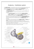
The vestibular labyrinth is a bony cavity located within the petrous portion of the temporal
bone → it consists of the 3 semicircular canals, 2 otolithic organs (utricle and saccule) and the
cochlea (= part of the hearing apparatus)
- Semicircular canals: 3 membranous channels located within the bony semicircular
ducts of the labyrinth filled with endolymph
o They are located in 3 planes, each plane making an angle of approximately 90
degrees with the other
▪ Anterior (superior) → sagittal plane
▪ Lateral (horizontal) → transverse plane
▪ Posterior (inferior) → frontal plane
o The terminal part of each canal ends with a dilation called the ampulla, which
opens into the vestibule
▪ The ampulla of each semicircular canal contains a cluster of
mechanoreceptor cells called the crista ampullaris
▪ Each crista is composed of the special sensory receptor cells, called the
hair cells
o The movement of the endolymph stimulates the hair cells → this way, each
semicircular canal detects when the head moves during the rotational
acceleration along its corresponding plane → thus it can detect head
movements such as nodding up/down, shaking side to side, tilting left/right


Figure 1: Adapted from a lecture of Raymond van de Berg

- Otolithic organs: 2 membranous cavities that lie in the bony vestibule of the inner ear
o Utricle: this lies in the posterior part of the vestibule
▪ On one end, it communicates with the semicircular canals, whilst on
the opposite end it forms a utriculosaccular duct with the saccule
▪ This duct passes through the temporal bone and reaches the posterior
surface of the petrous part of the temporal bone


@MyMedicalPulse

The benefits of buying summaries with Stuvia:

Guaranteed quality through customer reviews

Guaranteed quality through customer reviews

Stuvia customers have reviewed more than 700,000 summaries. This how you know that you are buying the best documents.

Quick and easy check-out

Quick and easy check-out

You can quickly pay through credit card or Stuvia-credit for the summaries. There is no membership needed.

Focus on what matters

Focus on what matters

Your fellow students write the study notes themselves, which is why the documents are always reliable and up-to-date. This ensures you quickly get to the core!

Frequently asked questions

What do I get when I buy this document?

You get a PDF, available immediately after your purchase. The purchased document is accessible anytime, anywhere and indefinitely through your profile.

Satisfaction guarantee: how does it work?

Our satisfaction guarantee ensures that you always find a study document that suits you well. You fill out a form, and our customer service team takes care of the rest.

Who am I buying these notes from?

Stuvia is a marketplace, so you are not buying this document from us, but from seller mymedicalpulse. Stuvia facilitates payment to the seller.

Will I be stuck with a subscription?

No, you only buy these notes for $0.00. You're not tied to anything after your purchase.

Can Stuvia be trusted?

4.6 stars on Google & Trustpilot (+1000 reviews)

65507 documents were sold in the last 30 days

Founded in 2010, the go-to place to buy study notes for 15 years now

Start selling
Free  1x  sold
  • (0)