100% satisfaction guarantee Immediately available after payment Both online and in PDF No strings attached
logo-home
Summary Neural Basis of Cognition and Perception $7.49   Add to cart

Summary

Summary Neural Basis of Cognition and Perception

1 review
 125 views  17 purchases
  • Course
  • Institution
  • Book

Summary of all the lectures covered for this course, including extra research done on some topics for an in-depth knowledge about the topics. The summary includes informative text as well as supporting figures. With this summary you can prepare for your exam in an easy and understandable manner whi...

[Show more]

Preview 4 out of 31  pages

  • No
  • 15,17,18, 19-29, 30-32, 52-54, 65-67
  • March 19, 2022
  • 31
  • 2021/2022
  • Summary

1  review

review-writer-avatar

By: arlumoindong • 2 year ago

avatar-seller
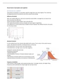
Neural basis of perception and cognition

Somatosensory system
The function of sensation is to transform specific energy forms into neural signals. This is done by:
De/Hyperpolarization (transduction) and action potential (encoding).

Difference threshold

JND= just notable difference. How much should two stimuli differ in strength for our senses to be
able to perceive the difference.
Fechner was able to explain Weber’s law to describe this:
•The relationship between stimulus strength and neural signal is logarithmic
•The difference in neural activity that is needed to discern between two different stimuli is the same
for all stimulus levels.


Absolute threshold

It is the smallest level of an stimulus detectable by the senses. Due to noise in the central nervous
system, the absolute threshold is not determined in a sharp way.
A threshold that is too low causes more false positives
A threshold that is too high causes more false negatives.

A design for stimulus detection task:
Distributions (Gaussian)
-Standard deviations
-Discriminability is function of distance between peaks and
overlap
-Ideal observer minimizes total errors and maximizes correct
responses (decision boundary at intersection of distributions)


Frequency modulation
If the stimulus is high enough, action potential takes place. A stronger stimulus, the higher firing
frequency (action potentials do not get larger but you have more).

,Somatosensory system
The somatosensory system has pathways of:
–Tactile reception: detection of shape and size of objects
–Nociception: detection of pain
–Thermoception: detection of heat and cold

Dorsal root ganglia (DRG) have cell bodies outside spinal cord. Information from the receptor is
conveyed by action potential.


Transduction takes place when there is enough stimuli for action potential to be triggered. Channels
open up (transduction) in different way:
-Lipid tension (osmotic swelling)
-Structural proteins linked to ion channel
-Indirect: forces conveyed to a force sensor

Skin receptors
Mechanoreceptors encapsulate afferent fibres and turn them to specific features of somatic
stimulation (myelinated). Unmyelinated fibres with free nerve endings are for temperature and pain.
Every receptor has its own receptive field. Receptive field of a sensory neuron is the spatial domain in
the sense organ where stimulation excites or inhibits the neuron. This is where you can measure the
action potential by a recording electrode and you will hear the spikes.

Minimum interstimulus distance is required to perceive two simultaneous stimuli as distinct. This is
related to the:
•Two-point discrimination threshold
•Receptive field size
•Periphery receptor density
•Cortical magnification factor

Sensory integration from multiple receptors
There are neurons that are excited by pressing the receptive field and neurons that are inhibited.
This lateral inhibition by local interneurons confines excitation to the central zone, enhancing the
contrast/sensitivity to changes.

,Tactile reception = Detection of shape and sizes of objects by mechanoreceptors.
Haptics=active touching for interpretation of spatiotemporal patterns of stimuli.
Big and small receptive fields are related to the depth in the skin (detection of coarse and fine
textures) Slow and fast adapting is the specialization in low and high temporal frequencies. Different
receptive fields are used for different tasks: Meisner (force rate, movement), Merkel (grip force),
Pacinian (vibrations at contact), Ruffini (hand posture).

Nociception= Detection of tissue damage (sensed as pain).
Free nerve endings are involved and nociceptors do not respond to light touch. First and second pain
stimuli are carried by two different primary afferent fibres. The propagation speed is a function of
the diameter and depends on axon myelinization.

Hyperalgesia: following painful stimulus, stimuli around the injured area are perceived as more
painful. Exp. Sunburn.

Different pathways:


Thermoreception= Detection of heat and cold
Two groups of sensors (free nerve endings): warmth (unmyelinated C fibers) cold receptors (C &
myelinated Aδ fibers)
There are opposite responses to warm and cold stimuli and this stimuli is strongly adapting.

Somatosensory cortex

Brodmann’s areas: VPM: VPL:
S3a: Proprioception face from body & posterior head
S3b & S1: cutaneous
S2: both

The somatosensory cortex has a columnar organization and is organized on the type of input that you
get and with respect to the size of the receptive field. So the type of somatosensory information and
size of the receptive fields differ amongst the different areas of somatosensory cortex.

, Sensory input that is particularly significant to a given species gets relatively more cortical
representation. For us the fingers have a large cortical representation.

The neurons don’t only respond to touch but there is a Motion and orientation selective responses in
S2. Decreased activation in S2 takes place when the subject is distracted by a visual task or the
stimuli is repetitively stimulated.
The somatosensory cortex has plasticity. Functional cortical remapping takes place by changes in the
somatosensory map after amputation of a digit or fusion of a digit. It also takes place after repetitive
use of digits.


This also happens for the whiskers of rodents. If one whisker falls of by damage, the other parts of
the somatosensory cortex grow larger to compensate for the loss of the whisker.

Panthom pain
Cortical remapping could be an explanation for phantom pain. After amputation, most patients feel
as though the missing organ is still present. This feeling diminishes over time, but can be reactivated
by disruptions to the stump. The cortical activation is expanded in patients with amputees. Mirror
therapy can help for panthom pain.
Signals from nociceptors in the viscera can be felt as “referred pain” elsewhere in the body
(e.g. Myocardial infarction) because neurons converge on same the projection.(figure below)


Mirror neurons and autism
Mirror neurons
Mirror neurons fire not only when an animal or human performs an action, but also when passively
observing others carrying out the same actions. (yawning) The neurons have a visible component and
motory component. So respond to vision and to ‘doing something’. They are found everywhere
including in corticospinal neurons. The function is suggested to be Involved in action understanding,
(Learning by) imitation, empathy; social cognition and disorders like autism.
It is possible to experimentally demonstrate the existence of mirror neurons. For example in an
experiment with a monkey grabbing a treat from your hand and recording brain activity. Next when
the monkey looks at your hand and you yourself grab the treat from your hand, the same brain
activity is recorded. It does not work when the human uses a tool to grab the treat. Response can be
dependent on gaze direction of actor. Meaning whether the actor is looking at the person and is
moving directed to the person or not while conducting the experiment.

The benefits of buying summaries with Stuvia:

Guaranteed quality through customer reviews

Guaranteed quality through customer reviews

Stuvia customers have reviewed more than 700,000 summaries. This how you know that you are buying the best documents.

Quick and easy check-out

Quick and easy check-out

You can quickly pay through credit card or Stuvia-credit for the summaries. There is no membership needed.

Focus on what matters

Focus on what matters

Your fellow students write the study notes themselves, which is why the documents are always reliable and up-to-date. This ensures you quickly get to the core!

Frequently asked questions

What do I get when I buy this document?

You get a PDF, available immediately after your purchase. The purchased document is accessible anytime, anywhere and indefinitely through your profile.

Satisfaction guarantee: how does it work?

Our satisfaction guarantee ensures that you always find a study document that suits you well. You fill out a form, and our customer service team takes care of the rest.

Who am I buying these notes from?

Stuvia is a marketplace, so you are not buying this document from us, but from seller StephSilentium. Stuvia facilitates payment to the seller.

Will I be stuck with a subscription?

No, you only buy these notes for $7.49. You're not tied to anything after your purchase.

Can Stuvia be trusted?

4.6 stars on Google & Trustpilot (+1000 reviews)

57114 documents were sold in the last 30 days

Founded in 2010, the go-to place to buy study notes for 14 years now

Start selling
$7.49  17x  sold
  • (1)
  Add to cart