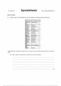
▪ Components of the respiratory system ;
Nose → Pharynx → Larynx → Trachea → Bronchi → Lung
Upper Respiratory Lower Respiratory
System System
▪ 2 Functional components of the respiratory system ;
1) The conducting portion - Nasal cavity , pharynx , larynx , trachea , bronchi , bronchioles , terminal bronchioles
* Functions :- to warm, humidify and clean inhaled air
2) The respiratory portion - Respiratory bronchioles , alveolar ducts , alveolar sac , alveoli
* Function :- to exchange gas
Trachea - 3 layers
1. Mucosa
1) Epithelium - Pseudostratified ciliated cells and mucous (goblet) cells are the two major components of the
epithelium.
• Clear basement membrane
• Pseudostratified ciliated columnar epithelium contains ;
a) Ciliated cells : columnar with cilia
b) Goblet cells
c) Basal cells : pyramidal shape , basally-located , undifferentiated cells, can differentiate into ciliated
cell or goblet cell
d) Brush cells : columnar , microvilli , EM – RER , no granules.
* Function - become into ciliated cell and receive sensory stimuli
e) Small granule cells : less , pyramidal and EM: dense-core granules, in neuroepithelial body.
* Function - secret hormones to regulate contract of SM and secretion of gland
✓ 5-hydroxytryptamine
✓ Calcitonin
2) Lamina propria (loose CT) - contain LC , PC , MC , BV , LV
3) Smooth muscle
2. Submucosa - LCT with blood vessels, lymphatic vessels and nodes with Tracheal glands, mixed glands
3. Adventitia - 16-20 C - shaped cartilage rings and a Circular ligament composed of elastic fibers
* In the posterior part of the trachea (membrane part) , the adventitia contains smooth muscle , elastic fiber ,
tracheal glands
1
, ▪ The trachea is lined with typical respiratory mucosa in which the lamina propria contains numerous seromucous
glands producing watery mucus .
▪ A series with about a dozen C-shaped rings of hyaline cartilage between the submucosa and adventitia reinforces
the wall and keeps the tracheal lumen open .
▪ The open ends of the cartilage rings are on the posterior surface , against the esophagus and are bridged by a
bundle of smooth muscle called the trachealis muscle and a sheet of fibroelastic tissue attached to the
perichondrium.
General structure of Lungs
1) Capsule - visceral layer of pleura-serous membrane mesothelium and connective tissue
2) Parenchyma - all branches of bronchi and alveoli ( right 3 , left 2 )
3) Interstitial substance - connective tissue , blood vessels , lymphatic vessels , nerves
▪ The pulmonary lobules are each pyramid-shaped , with the apex aimed at the pulmonary hilum and each is
delineated by a thin layer of connective tissue , which in adults is frequently incomplete.
▪ Moving through the smaller bronchi and bronchioles toward the respiratory portion, the histological organization of
both the epithelium and the underlying lamina propria gradually becomes more simplified.
▪ The trachea divides into two primary bronchi that enter each lung at the hilum , along with arteries , veins and
lymphatic vessels.
▪ After entering the lungs , the primary bronchi course downward and outward , giving rise to three secondary (lobar)
bronchi in the right lung and two in the left lung , each of which supplies a pulmonary lobe.
▪ These lobar bronchi again divide , forming tertiary (segmental) bronchi.
▪ Each of the tertiary bronchi , together with the smaller branches it supplies , constitutes a bronchopulmonary
segment —with its own connective tissue capsule and blood supply.
▪ The existence of such lung segments facilitates the specific surgical resection of diseased lung tissue without
affecting nearby healthy tissue.
▪ The tertiary bronchi give rise to smaller and smaller bronchi, whose terminal branches are called bronchioles.
▪ Each bronchiole enters a pulmonary lobule , where it branches to form five to seven terminal bronchioles.
The conducting portion - Lobar bronchi to small bronchi
1. Mucosa
a) respiratory epithelium
b) lamina propria : thinner, SM [ contains crisscrossing bundles of spirally arranged smooth muscle and elastic
fibers ]
2. Submucosa : mixed glands
3. Adventitia : cartilage plate
2