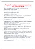
usmle step 1 questions and answers with all correct answers 2022
what gland is found in the muscular triangle of the neck
hat is the name of the spinal cord passing within the subarachnoid spac
Connected book
Book Title:
Author(s):
Edition:
ISBN:
Edition:
Written for
USMLE
All documents for this subject (1213)
1
review
By: praveenkumar8 • 1 year ago
By: Nipsey • 1 year ago
Thanks for the review & success in your exam
Seller
Follow
Nipsey
Reviews received
Content preview
USMLE Step 1 Questions And Answers With All
Correct Answers 2022/2023
What gland is found in the muscular triangle of the neck? Correct Answer: Thyroid gland
Is an afferent or efferent pupillary defect described as B/L pupillary constriction when light is shined in
the unaffected eye and B/L paradoxical dilation when light is shined in the affected eye? Correct
Answer: Afferent pupillary defect (CN II lesion); in an efferent pupillary defect (CN III), B/L constrict
when light is shined in the unaffected eye and consensual pupil constriction occurs when light is
shined in the affected eye.
What is the name of the spinal cord passing within the subarachnoid space and forming the spinal
nerves that exit the lumbar and sacral foramina? Correct Answer: Cauda equine
Name the laryngeal muscle described by the following: • Pulls the arytenoids cartilages closer to the
thyroid, relaxing the vocal ligaments and thereby decreasing the pitch Correct Answer:
Thyroarytenoid muscles
Name the laryngeal muscle described by the following: • Tenses the vocal ligaments, increasing the
distance between the cartilages, thereby increasing the pitch Correct Answer: Cricothyroid muscles
Name the laryngeal muscle described by the following: • adducts the vocal ligaments, closes the air
passageway during swallowing, and allows phonation Correct Answer: Lateral cricoarytenoid muscles
Name the laryngeal muscle described by the following: • Only muscle to abduct the vocal cords
Correct Answer: Posterior cricoarytenoid muscles
Where does the parotid (Stensen's) duct enter the oral cavity? Correct Answer: Opposite the second
upper molar tooth
From what aortic arch are the following structures derived? • Common and internal carotid arteries
Correct Answer: Third aortic arch MS CARD is my mnemonic for the aortic arch derivatives
From what aortic arch are the following structures derived? • Degenerates Correct Answer: Fifth MS
CARD is my mnemonic for the aortic arch derivatives
From what aortic arch are the following structures derived? • Stapes artery Correct Answer: Second
MS CARD is my mnemonic for the aortic arch derivatives
From what aortic arch are the following structures derived? • Maxillary artery Correct Answer: First
MS CARD is my mnemonic for the aortic arch derivatives
From what aortic arch are the following structures derived? • Arch of the aorta and right subclavian
artery Correct Answer: Fourth MS CARD is my mnemonic for the aortic arch derivatives
From what aortic arch are the following structures derived? • Right and left pulmonary arteries and
the ductus arteriosus Correct Answer: Sixth MS CARD is my mnemonic for the aortic arch derivatives
,What abdominal muscle contributes to the anterior layer of the rectus sheath, forms the inguinal
ligament, and in men gives rise to the external spermatic fascia of the spermatic cord? Correct
Answer: External abdominal oblique
Name the compartment of the lower extremity and the nerve based on its movements. • Adduct the
thigh and flex the hip Correct Answer: Medial compartment of the thigh, obturator nerve
Name the compartment of the lower extremity and the nerve based on its movements. • Plantar flex
the foot, flex the toes, and invert the foot Correct Answer: Posterior compartment of the leg, tibial
nerve
Name the compartment of the lower extremity and the nerve based on its movements. • Dorsiflex the
foot, extend the toes, and invert the foot Correct Answer: Anterior compartment of the leg, deep
peroneal nerve
Name the compartment of the lower extremity and the nerve based on its movements. • Flex the hip
and extend the knee Correct Answer: Anterior compartment of the thigh, femoral nerve
Name the compartment of the lower extremity and the nerve based on its movements. • Extend the
hip and flex the knee Correct Answer: Posterior compartment of the thigh, tibial nerve
Name the compartment of the lower extremity and the nerve based on its movements. • Plantar flex
the foot and evert the foot Correct Answer: Lateral compartment of the leg, superficial peroneal nerve
What are the five branches of the posterior cord of the brachial plexus? Correct Answer: STARS 1.
Upper Subscapularis 2. Thoracodorsal 3. Axillary 4. Radial 5. Lower Subscapularis
Name the correct artery. • The right recurrent laryngeal nerve passes around it. Correct Answer:
Right brachiocephalic artery
Name the correct artery. • The left recurrent laryngeal nerve passes around it. Correct Answer: Arch
of the aorta
The inferior mesenteric artery drains into it. Correct Answer: The splenic vein
Are the quadrate and caudate lobes of the liver functionally part of the left or right lobe? Correct
Answer: Functionally they are part of the left lobe of the liver because they receive their blood supply
from the left hepatic artery. Anatomically they are considered part of the right lobe of the liver.
What bones make up the acetabulum? Correct Answer: Pubis, ilium, and ischium
What is the anatomic positioning of the right and left gastric nerve plexus of the esophagus as they
pass through the diaphragm? Correct Answer: LARP: Left goes Anterior and Right goes Posterior
(because of the rotation of the gut; remember your embryology!)
What vessel is lacerated in an epidural hematoma? Correct Answer: Middle meningeal artery
True or false? Below the arcuate line, all the aponeurotic fibers run anterior to the rectus abdominis.
Correct Answer: TRUE
,What ocular muscle • Adducts the eyeball and is involved in horizontal conjugate gaze? Correct
Answer: Medial rectus (CN III) (LR6 SO4)3
What ocular muscle • Elevates and adducts the eyeball? Correct Answer: Superior rectus (CN III)
(LR6 SO4)3
What ocular muscle • Depresses and abducts the eyeball? Correct Answer: Superior Oblique (CN IV)
(LR6 SO4)3
What ocular muscle • Elevates and abducts the eyeball? Correct Answer: Inferior Oblique (CN III)
(LR6 SO4)3
What ocular muscle • Abducts the eyeball and is involved in horizontal conjugate gaze? Correct
Answer: Lateral rectus (CN VI) (LR6 SO4)3
What ocular muscle • Depresses and adducts the eyeball? Correct Answer: Inferior rectus (CN III)
(LR6 SO4)3
Which muscles of the eye are under parasympathetic control? Correct Answer: Constrictor pupillae
and ciliary muscles
Which direction does the uvula deviate in a left vagus nerve lesion? Correct Answer: A left CN X
lesion results in the uvula deviating to the right. (Uvula points away from the affected side.)
Is a subdural hematoma an arterial or venous bleed? Correct Answer: Subdural hematoma is a
rupture of the cerebral veins where they enter the superior sagittal sinus.
Which CNs are found in the midline of the brainstem? Correct Answer: CN I, II, III, VI, and XII Add 1 +
1 = 2, 1 + 2 = 3, 1 + 2 + 3 = 6, 1 + 2 + 3 + 6 = 12
What muscles insert in or on the intertubercular groove of the humerus? Correct Answer: "Lady
between two Majors": latissimus dorsi, pectoralis major, and teres major
What nerve supplies taste sensation to the anterior two-thirds of the tongue? Correct Answer: Chorda
tympani of CN VII
What part of the heart forms • The right border? Correct Answer: Right atrium
What part of the heart forms • Left border? Correct Answer: Left ventricle and auricle of left atrium
What part of the heart forms • Apex? Correct Answer: Tip of the left ventricle
What part of the heart forms • Base? Correct Answer: Left atrium and tip of the right atrium
What part of the heart forms • Superior border? Correct Answer: Conus arteriosus of the right
ventricle and right and left auricles
What part of the heart forms • Anterior wall? Correct Answer: Right ventricle
What part of the heart forms • Posterior wall? Correct Answer: Left atrium
, What part of the heart forms • Diaphragmatic wall? Correct Answer: Left ventricle and tip of right
ventricle
What nerves carry the sensory and motor components of the blink reflex? Correct Answer: CN V1
carries the sensory and CN VII carries the motor component of the blink reflex.
What muscle keeps the stapes taut against the oval window? Correct Answer: Stapedius muscle
Name the components of the femoral canal, working laterally to medially. Correct Answer: NAVEL:
Femoral Nerve, Artery, Vein, Empty space, and Lymphatics/Lacunar ligament
What muscle is most superior in the orbit? Correct Answer: Levator palpebrae superioris
What portion of the pericardium adheres to the tunica adventitia of the great vessels? Correct
Answer: Fibrous pericardium
What two veins form the portal vein? Correct Answer: The superior mesenteric vein and the splenic
vein (after it receives the inferior mesenteric vein) join to form the portal vein.
What CNs are responsible for the sensor and motor components of the light reflex? Correct Answer:
CN II is the sensory limb and CN III is the motor component through parasympathetic stimulation.
Arrange the following layers in the correct sequence through which a needle must pass in a lumbar
puncture. • Skin • Subarachnoid space • Interspinous ligament • Dura mater • Deep fascia • Epidural
space • Superficial fascia • Interlaminar space • Supraspinous ligament • Arachnoid mater Correct
Answer: During a lumbar puncture the needle passes through the interlaminar space in the midline of
L3-L4, with the tip of the iliac crest in the flexed position as the landmark. Order of puncture: 1. Skin
2. Superficial fascia 3. Deep fascia 4. Supraspinous ligament 5. Interspinous ligament 6. Interlaminar
space 7. Epidural space 8. Dura mater 9. Arachnoid mater 10. Subarachnoid space. (They ask this in
some variation every year, so know it.)
What ocular ganglion is affected if the pupil on the affected side sluggishly responds to light with
normal accommodation? Correct Answer: Ciliary ganglion producing a tonic pupil
What is the name for the most prominent spinous process? Correct Answer: Vertebra prominens (C7
in 70% of cases, C6 in 20%, T1 in 10%)
What muscles make up the rotator cuff? Correct Answer: SITS—Subscapularis, Infraspinatus, Teres
minor, Supraspinatus
What is the function of white rami communicantes? Correct Answer: They are preganglionic
sympathetic axons. They are white because they are myelinated.
What muscle or muscles are innervated by the following nerves? • Suprascapular nerve Correct
Answer: Supraspinatus and infraspinatus
What muscle or muscles are innervated by the following nerves? • Upper subscapularis nerve Correct
Answer: Subscapularis
The benefits of buying summaries with Stuvia:
Guaranteed quality through customer reviews
Stuvia customers have reviewed more than 700,000 summaries. This how you know that you are buying the best documents.
Quick and easy check-out
You can quickly pay through credit card or Stuvia-credit for the summaries. There is no membership needed.
Focus on what matters
Your fellow students write the study notes themselves, which is why the documents are always reliable and up-to-date. This ensures you quickly get to the core!
Frequently asked questions
What do I get when I buy this document?
You get a PDF, available immediately after your purchase. The purchased document is accessible anytime, anywhere and indefinitely through your profile.
Satisfaction guarantee: how does it work?
Our satisfaction guarantee ensures that you always find a study document that suits you well. You fill out a form, and our customer service team takes care of the rest.
Who am I buying these notes from?
Stuvia is a marketplace, so you are not buying this document from us, but from seller Nipsey. Stuvia facilitates payment to the seller.
Will I be stuck with a subscription?
No, you only buy these notes for $16.99. You're not tied to anything after your purchase.