Test Bank –Fundamentals Of Anatomy & Physiology, by Frederic Martini , Judi Nath ,Edwin Bartholomew 12th Edition||ALL NEW 2024
Histology exam preparation with a Summary of Fundamentals of Anatomy & Physiology, Global Edition - Cell Biology Histology
Test Bank Martini, Nath, Bartholomew Fundamentals of Anatomy & Physiology, Global Edition, 12th Edition Latest Verified Review 2024 Practice Questions and Answers for Exam Preparation, 100% Correct wi...
All for this textbook (28)
Written for
Vrije Universiteit Amsterdam (VU)
Bachelor Biomedical Sciences
Human Anatomy And Physiology (AB_1197)
All documents for this subject (8)
Seller
Follow
lente90
Reviews received
Content preview
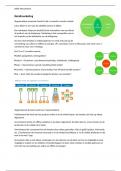
Physiology of the heart
Pulmonary circulation (= only lungs) and systemic circulation (=whole
body). If there are no separate circulations, always everything low
pressure. Relaxation needed to let new blood in. contraction
following electrical stimulation of cardiomyocytes. Automation of the
heart by pacemaker cells.
Conduction
Pacemaker cells are located in SA node on top of right atrium. The
action potential in the heart goes from SA node cell to ventricular cell. They have different action
potentials (different sodium channel):
Pacemaker cells unstable resting potential and slow depolarization prepotential
o Sodium going in makes membrane potential go up to pass threshold calcium
channels also open potassium opens later and goes out again
Ventricular cells need a signal by reacting to environment and quick depolarization
Spread action potential
Potential goes from SA node to AV node, this gives a delay while atrial contraction takes place.
The AV bundle conducts to the bundle branches (left bundle branch is larger) and conduct to
Purkinje fibers, through moderator band to papillary muscles in right ventricle. Because of this the
papillary muscles contract before the ventricles already closing the valves. The Purkinje fibers start
contraction at the apex spreading towards the base.
Calcium entering the cell is not enough to activate the full muscle, sarcoplasmic reticulum releases
more calcium upon entering RyR (=CICR), binding calcium to myofilament initiates contraction. For
relaxation SERCA pumps calcium back in SR. ATP is used for relaxation, not contraction, if there is no
ATP only contraction.
Electrocardiogram
ECG reflects electrical differences between regions in the heart. Meaning when none of
the cells are depolarized or all of the cells are depolarized there is no signal measured.
1. P wave = begin none of cells depolarized, end all of them depolarized of SA node
2. QRS-complex = signal through AV node spreading through ventricles, ventricle
depolarization and atrial repolarization
3. T wave = repolarization of ventricles
Location of electrodes affect the way you look at the heart. Size of the heart also affects the
depolarization wave and distance of the electrodes to the heart. There are fixed pairs of
electrodes to measure in the right directions Einthoven’s triangle.
Abnormalities
Lead one measures right to left, when there is an inversion in the heart, the ECG will show negative
spikes instead of positive. QRS – nothing – T wave does not occur in a heart attack coronary artery
blocked and no point with all cells repolarized. Ectopic beats are an extra systole in the rhythm,
shows an extra QRS. This can be in the atria (=supraventricular) or in the ventricles (=ventricular).
,Cardiac cycle
Blood flow through the heart is determined by pressure differences. VA valves are: tricuspid (=right)
and mitral (=left).
Passive filling = AV valves open, aorta/pulmonary valve closed
Atrial kick = AV valves open, aorta/pulmonary valve closed
Isovolumetric contraction = AV valves closed, aorta/pulmonary valve closed, not yet ejection
Ejection = AV valves closed, aorta/pulmonary valve open, blood goes out
Isovolumetric relaxation = AV valves closed, aorta/pulmonary valve closed
The pumping of the heart makes 2 sounds. The first sound is caused by closing of the tricuspid and
mitral valve because of the systole (low pressure and low frequency), Ventricular contraction. The
second sound is made by the closing of the aorta and pulmonary valves in diastole (high pressure
and high frequency), filling of ventricles. Third heart is associated with blood flowing into the
ventricles. Fourth heart sound of the atrial contraction.
Formulas
Stroke volume = end diastolic volume – end systolic volume
Ejection fraction = EDV – ESV / EDV
EDV = end diastolic volume, determined by available filling time and venous return
ESV = end systolic volume, determined by preload, contractility and afterload
Cardiac output (ml/min) = stroke volume X heart rate
adjustments
Affecting stroke volume can be done in 3 ways:
Frank-starling mechanism = increased filling pressure leads to increased stroke volume
Adrenergic stimulation = Contractility of heart muscle by sympathetic noradrenaline
Afterload = force needed to open aortic and pulmonary valve
o Increases when blood vessels vasoconstrict
o Decreases when blood vessels vasodilate
Long term high demand of blood hypertrophy of the ventricles, caused by exercise or pregnancy
or after myocardial infarction (same amount of work with less muscle).
Autonomic innervation
Cardiac plexus innervates with SA and AV nodes, more sympathetic fibers. Cardioacceleratory center
controls sympathetic neurons to increase heart rate. Cardioinhibitory center control
parasympathetic neurons to slow the heart rate. These centers react to changes in BP and arterial
concentrations. Without innervation the heart will beat 80 – 100 bpm, therefore mostly
parasympathetic innervation. Changing if the heart rate is done by changing ionic permeability of
cells. Heart rate determined by resting membrane potential of the SA node cells:
Sympathetic stimulation = using noradrenaline to open sodium and calcium channels
Parasympathetic stimulation = acetylcholine to open up potassium channels
, Lecture 2 Anatomy of the heart
From Sternal angle to the 5th intercostal space parasternal on the right and 5 th
intercostal space midclavicular on the left. The base of the heart sits at the level of the
third costal cartilage to the left. Mediastinum = area above diaphragm and below
thoracic inlet between left and right pleural cavity, from sternum to vertebrae:
Superior mediastinum = (yellow) vagal nerves (above), left recurrent
laryngeal nerve, phrenic nerves (supports diaphragm, to side of the heart),
sympathetic trunk (besides vertebrae).
Inferior mediastinum
o Anterior = (green) artery and veins thoracicae internae (next to thymus)
o Middle = blue
o Posterior = (red) vessels (azygos vein right side enters in vena cava superior),
esophagus, thoracic duct, nerves
pericardium
Pericardial cavity formed by pericardium heart is pressed up against
the inside. Consist of 2 layers:
Fibrous pericardium = outer side, collagen fibers stabilizing
heart
Serous pericardium = inside of 2 layers
o Parietal = outside of the heart,
o Visceral = inside touching the organ, epicardium
Pericardial fluid between the serous layers forms the pericardial
cavity. Infection in the pericardium can cause pericarditis
inflamed surfaces rubbing against each other causes friction rub
and excess pericardial fluid production. This collecting in the
cavity causes cardiac tamponade.
Heart
Brachiocephalic trunk is first split off of the aorta, later splitting
into an artery to the head and the arm. Left subclavian artery
goes to the other arm. Right ventricle is rotated to the front, the
pulmonary artery comes from this. Both atria have an auricle
(little ear) = expandable part. Coronary sulcus marks border between atria and ventricles. Anterior
and posterior interventricular sulcus mark the boundary between the ventricles. Posterior wall of
atria are smooth surfaces, the anterior wall contains pectinate muscle.
Bloodflow
From right atrium, through right ventricle to the lungs. Inferior,
superior vena cava and coronary sinus enter the atrium. Blood
goes through tricuspid valve, attached via chordae tendineae,
fibers originate at the papillary muscles from ventricle. Internal
surface of right ventricle contains trabeculae carneae = muscular
ridges moderator band gives stimulus for contraction papillary
muscles. Blood goes through the conus arteriosus through the
pulmonary valve into the arteries.
The benefits of buying summaries with Stuvia:
Guaranteed quality through customer reviews
Stuvia customers have reviewed more than 700,000 summaries. This how you know that you are buying the best documents.
Quick and easy check-out
You can quickly pay through credit card or Stuvia-credit for the summaries. There is no membership needed.
Focus on what matters
Your fellow students write the study notes themselves, which is why the documents are always reliable and up-to-date. This ensures you quickly get to the core!
Frequently asked questions
What do I get when I buy this document?
You get a PDF, available immediately after your purchase. The purchased document is accessible anytime, anywhere and indefinitely through your profile.
Satisfaction guarantee: how does it work?
Our satisfaction guarantee ensures that you always find a study document that suits you well. You fill out a form, and our customer service team takes care of the rest.
Who am I buying these notes from?
Stuvia is a marketplace, so you are not buying this document from us, but from seller lente90. Stuvia facilitates payment to the seller.
Will I be stuck with a subscription?
No, you only buy these notes for $6.84. You're not tied to anything after your purchase.