100% satisfaction guarantee Immediately available after payment Both online and in PDF No strings attached
logo-home
Pathology lecture notes $4.81
Add to cart

Class notes

Pathology lecture notes

 7 views  0 purchase
  • Course
  • Institution

This document includes a summary/ notes of all lectures throughout the course pathology, including practice questions + correct answers at the end of the document. These questions are used for previous exams and might help to understand the subjects.

Preview 4 out of 55  pages

  • December 25, 2022
  • 55
  • 2021/2022
  • Class notes
  • No name
  • All classes
avatar-seller
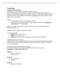
Pathology
Introduction into pathology
Pathology is the study of the causes and effects of disease or injury.
The word pathology also refers to the study of disease in general, incorporating a wide range of
biology research fields and medical practices. However, when used in the context of modern medical
treatment, the term is often referred to the diagnose of diseases, mostly through analysis of organs,
tissues, cells, and body fluids.

Disease
 Any abnormality that causes loss of health (‘ill health’).
 Characterized by a specific set of features (signs, symptoms, functional and morphological
manifestations) that are ‘not normal’.

What does ‘normal’ mean? Normal is the most frequent state in a population defined by age
distribution, gender, etc.

Pathology is part of a disease’s systematic description:

Technical terminologies:
 Prefixes (hyper-, meta-, hypo-, etc.)
 Suffixes (-itis, -oma, -oid, etc.)
 Eponyms (e.g. name of person that described a disease, like Hopkin’s disease)

Pathology does not have a black box.
Diagnosis with the help of histology (biopsies, resections, frozen sections) and cytology (body fluids).
Looking under the microscope is not enough anymore for diagnosis, gene expression and genetics
etc. are also important.

Specimen grossing is performed in the cutting room.
You harden the tissue by embedding (with paraffin) to be able to cut it and stain it:
paraffin block > cutting > slides > H&E stain.

Diagnosis
 Benign vs. malignant?
 Type of tumor?

Prognosis
 TNM classification?
 Radicality?

Prediction
 Response to treatment?

,EXAM PART 1
Cell- and tissue adaptation, & damage
Ch2: Cell injury, cell death, and adaptations, pages 31-56

What is disease?
 A dysfunction of an organ or tissue, because of damage to the cells.
 The damage can be of many causes, chemical, thermal, radiation, DNA damage, micro
bacterial, etc.
 The damaging agent is the etiology, the influence on and the changes in cellular processes
are called the pathogenesis.

Missense mutation (e.g. sickle cell anemia); if you change even one molecule, it can change the
amino acid, and change the entire protein.

Internal milieu is optimised, and thus also attractive for intruders. Effective defence is required
 Infectious diseases.

Organisation and clear vision of tasks is mandatory, incl. discipline of cells, a.o. with regards to
proliferation
 Cancer.

Cell damage, stress and stressors
- Disease is caused by damages to (part of) a cell or group of cells (etiology).
- If the initial damage causes further damage you speak of pathogenesis.
- The cell/ organ reacts to minimize the impacts of damage (adaptation).
- Most of the damage that is done to cells is reversible, lead to adaptation or, ultimately to
death of the cell.

Reversible cell injury: cell swelling, fatty change, plasma membrane blebbing and loss of microvilli,
mitochondrial swelling, dilation of the ER, eosinophilia (due to decreased cytoplasmic RNA).
Ischemia (reduced blood supply) injures tissues faster and usually more severe than does hypoxia.

- Neutrophils: acute inflammatory response.
- Lymphocytes: at the end of an immune response. Prevent reactions against the body’s own
tissues. Lymphocytes are the mediators of adaptive immunity and the only cells that produce
specific and diverse receptors for antigens.
- T-lymphocytes express antigen receptors called T-cell receptors (TCRs) that recognize
peptide fragments of protein antigens that are displayed by MHC molecules on the surface of
antigen-presenting cells.
- B-lymphocytes express membrane bound antibodies that recognize a wide variety of
antigens. B cells are activated to become plasma cells, which secrete antibodies.
- Cytotoxic T-lymphocytes: a defence mechanism against viruses and tumors that serves to kill
virus-infected and neoplastic cells (by directly inducing the effector phase of apoptosis).
- Natural killer cells kill cells that are infected by some microbes or are stressed and damaged
beyond repair. NK cells express inhibitory receptors that recognize MHC molecules that are
normally expressed on healthy cells and are thus prevented from killing normal cells.
- APCs capture microbes and other antigens, transport them to lymphoid organs, and display
them for recognition by lymphocytes.

Some forms of adaptation:

,1. Hypertrophy is the adaptation to increased workload (e.g. adapted myocyte: increase in size, but
not in numbers of cells!).
2. Hyperplasia is an increase in the number (not in the size!) of cells (e.g. breast feeding).
Uterus both hypertrophy and hyperplasia?
3. Atrophy is the decrease of tissue by decrease of cells size and/ or number (e.g. brain of elderly).
 Proteasomal degradation: destroying of proteins.
 Autophagy: in poor conditions of nutrition cell gets rid of a part of itself by eating parts up.
 Apoptosis: getting rid of cells?
4. Metaplasia (self-defence) is the replacement of one normal tissue by another (normal) tissue (e.g.
squamous metaplasia: columnar epithelium replaced by thicker squamous epithelial after smoking
or inhaling toxins).
If you stop smoking, does this change in epithelium reverse back to columnar epithelium?

Cell damage by oxygen shortage: the cell swells. Cause: failure of Na +-K+-ATPase pump.
 Necrosis: usually a group of cells, less staining because it is watery (swelling and eventually
falls apart, inflammatory response, always pathological). Extra info: increased eosinophilia;
nuclear shrinkage, fragmentation and dissolution; breakdown of plasma membrane and
organellar membranes; abundant myelin figures; leakage and enzymatic digestion of cellular
contents.
o If the organ is still intact but a part is missing (no nuclei > death), we speak of
coagulation necrosis. Typically caused by infarction, underlying tissue is preserved
for several days after death of cells .
o You also have colliquative necrosis, e.g. hole in the brain (tissue is cleared away),
which results in a transformation of the tissue into a liquid viscous mass.
o Caseous necrosis: bacteria cause severe damage to the surrounding the tissues,
cheese-like appearance (specific for tuberculosis).
o Fat necrosis in pancreatitis, white spots (calcium), enzymes released in the
surrounding area which is digested now.
o Fibrinoid necrosis a specific pattern of irreversible, uncontrolled cell death (ONLY in
arterial wall). Blood vessels are inflamed, the necrotic tissue resembles fibrin (hence
the name).
 Apoptosis: cell shrinks instead of swelling, dark staining because of the smaller space (occurs
continuously, anti-inflammatory response, part of normal physiology).
o Regulated mechanism of cell death that serves to eliminate unwanted and
irreparably damaged cells, with the least possible host reaction, characterized by
enzymatic degradation of proteins and DNA, initiated by caspases; and by rapid
recognition and removal of dead cells by phagocytes.

, Know which cell does what?

Cell injury: Growth factor withdrawal, DNA damage

Extrinsic apoptosis-induction
A lethal signal from outside the cell (FasL, TNF) triggers a cascade that leads to apoptosis through
receptor activation.

Mitochondrial intrinsic pathway of apoptosis-induction: comes through the mitochondria.
Lack of survival signals, or damage or stress of the cell itself, induces apoptosis.

BCL-2 and B-cell (follicular) lymphoma
Loss of apoptosis-response by overexpression of BCL2-gen (anti-apoptosis cell).
Cause: BCL2-gen is, by translocation, brought under control of the promotor of the heavy chain of
the immunoglobulin.

Cytochrome c functions in electron transport in oxidative phosphorylation and induces apoptosis.

The apoptosome activates caspase-9
1. Cytochrome C binds to Apaf-1 (apoptosis-activating factor-1)
2. The thus formed heptamere complex binds procaspase-9 and ATP
3. Procaspase-9 is activated and the cascade van caspase-activations ensues


If the unfolded protein response fails, the cell will go into apoptosis.

In apoptosis phosphatidylserine ‘flops’ to the external side of the cell membrane. This ‘facing
outwards’ of the phosphatidylserine indicates that the cell is undergoing apoptosis.
Necroptosis: no necrosis, no apoptosis.

The benefits of buying summaries with Stuvia:

Guaranteed quality through customer reviews

Guaranteed quality through customer reviews

Stuvia customers have reviewed more than 700,000 summaries. This how you know that you are buying the best documents.

Quick and easy check-out

Quick and easy check-out

You can quickly pay through credit card or Stuvia-credit for the summaries. There is no membership needed.

Focus on what matters

Focus on what matters

Your fellow students write the study notes themselves, which is why the documents are always reliable and up-to-date. This ensures you quickly get to the core!

Frequently asked questions

What do I get when I buy this document?

You get a PDF, available immediately after your purchase. The purchased document is accessible anytime, anywhere and indefinitely through your profile.

Satisfaction guarantee: how does it work?

Our satisfaction guarantee ensures that you always find a study document that suits you well. You fill out a form, and our customer service team takes care of the rest.

Who am I buying these notes from?

Stuvia is a marketplace, so you are not buying this document from us, but from seller arzuburak. Stuvia facilitates payment to the seller.

Will I be stuck with a subscription?

No, you only buy these notes for $4.81. You're not tied to anything after your purchase.

Can Stuvia be trusted?

4.6 stars on Google & Trustpilot (+1000 reviews)

53068 documents were sold in the last 30 days

Founded in 2010, the go-to place to buy study notes for 14 years now

Start selling
$4.81
  • (0)
Add to cart
Added