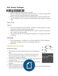
DNA Analysis Techniques
Separates DNA molecules on basis on length
DNA is a negatively charged molecule at neutral pH – in an electric charge, DNA
molecule will move towards the positive charge (anode)
Process: separating substances and analysing molecular structure based on the
rate of movement of each component in liquid medium under influence of electric
field
Types of gel
Agarose
Polysaccharide (derived from seaweed) – suitable for coarse separation of quite
large pieces of DNA (relatively low resolution)
Agarose powder mixed with TAE/TBE buffer – heated to molten then allowed to
set + nucleic acid stain (eg. EtBr)
Typically 1% used, 2% for finer separation/separation of fragments that differ
in a few bp
Acrylamide
Cross-linked polymer – suitable for very precise separation of DNA fragments
that differ in length by as little as 1 base (high resolution)
Neurotoxin
Visualising DNA (by staining)
Ethidium bromide
Flat molecule that intercalates between the stacked bases of
DNA
The orientation and proximity of ethidium with the stacked
bases causes the dye to display an increased fluorescence
compared to free dye (in solution)
UV radiation at 254nm is absorbed by the DNA and transmitted
to the bound dye
Energy is re-emitted at 590nm in the red-orange region of the
spectrum
Usually incorporated into the gel and running buffer
Stain is visualised by irradiating with a UV light source (ie. using a transilluminator)
and photographing with polaroid film