100% satisfaction guarantee Immediately available after payment Both online and in PDF No strings attached
logo-home
Samenvatting HC Lens, cataract $3.21
Add to cart

Summary

Samenvatting HC Lens, cataract

 7 views  0 purchase
  • Course
  • Institution

In deze samenvatting zijn de leerdoelen en aantekeningen van het hoorcollege verwerkt. Deze les is gegeven door Marc Veenstra, voor de studie Optometrie en Orthoptie, jaar 1.

Preview 2 out of 8  pages

  • February 22, 2023
  • 8
  • 2022/2023
  • Summary
avatar-seller
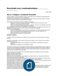
Hoorcollege 6 Lens, cataract

Leerdoelen

 de anatomie van de lens omschrijven

Zorgt ervoor dat het licht op de juiste manier wordt gebroken.


Cornea breekt rond de 43-48 dpt, de lens breekt rond de 20 dpt. En komt dan achter op het netvlies
terecht. Eerst buigt de cornea het licht naar binnen, daarna door de lens, en komt het beeld scherp
op de retina.

Als je jong bent gaat dit prima (geboorte 15 dpt, 25 jaar 8 dpt, ouder dan 50 1-2 dpt (presbyopie =
verziendheid door vermindering van het accommodatievermogen)), maar kom je boven de 50 jaar
dan gaat het systeem mindergoed werken.

De lens is dubbelbol (convex), doorzichtig,
9 mm groot.

De lens bestaat uit verschillende delen:
eptiheel, cortex, nucleus


De lens is opgebouwd uit drie delen:

, - Het kapsel (het zakje waar de lens in zit, en spant aan)
- Epitheel (bron van de lensvezels, zit een basaalmembraan, een barrière om grote
moleculen geen doorgang te laten vinden). Epitheel zit alleen aan de voorkant. De lens
ontstaat uit het ectoderm.
- De lensvezels zelf

Het kapsel is elastisch, zit veel elastine in, heeft snel de nijging om bol te gaan staan. Lens zit vast
met zonula vezels.

Oogbeker groeit tegen het ectoderm aan, en er ontstaat een hol bolletje en zakt het naar binnen en
ontstaat er uiteindelijk een lens.


Hoe dieper de lensvezels zitten, hoe ouder ze zijn. Ze differentiëren steeds.


Verschillende delen van de lens:

1. Kapsel (anterior)
2. Voorste lijn van de verdeling (tussen epitheel en cortex)
3. Voorkant van de volwassen nucleus
4. Voorkant van het oppervlak van de foetale nucleus
5. Voorste helft van embryonale nucleus (met daarin de Y structuur)
6. Achterste helft van embryonale nucleus (met daarin λ structuur)
7. Achterste oppervlak van foetale nucleus
8. Achterste oppervlak van volwassen nucleus
9. Achterste lijn (verdeling tussen cortex en kapsel)
10. Achterste kapsel


 het lenskapsel omschrijven

The benefits of buying summaries with Stuvia:

Guaranteed quality through customer reviews

Guaranteed quality through customer reviews

Stuvia customers have reviewed more than 700,000 summaries. This how you know that you are buying the best documents.

Quick and easy check-out

Quick and easy check-out

You can quickly pay through credit card or Stuvia-credit for the summaries. There is no membership needed.

Focus on what matters

Focus on what matters

Your fellow students write the study notes themselves, which is why the documents are always reliable and up-to-date. This ensures you quickly get to the core!

Frequently asked questions

What do I get when I buy this document?

You get a PDF, available immediately after your purchase. The purchased document is accessible anytime, anywhere and indefinitely through your profile.

Satisfaction guarantee: how does it work?

Our satisfaction guarantee ensures that you always find a study document that suits you well. You fill out a form, and our customer service team takes care of the rest.

Who am I buying these notes from?

Stuvia is a marketplace, so you are not buying this document from us, but from seller MiekeOrtho. Stuvia facilitates payment to the seller.

Will I be stuck with a subscription?

No, you only buy these notes for $3.21. You're not tied to anything after your purchase.

Can Stuvia be trusted?

4.6 stars on Google & Trustpilot (+1000 reviews)

52510 documents were sold in the last 30 days

Founded in 2010, the go-to place to buy study notes for 14 years now

Start selling
$3.21
  • (0)
Add to cart
Added