NBME 16 Block 1 and 2 (Answered) Verified Solution
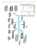
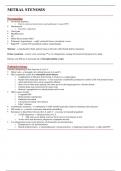
NBME 16 Block 1 and 2 (Answered) Verified Solution Lab values in Sarcoidosis 1.Elevated ACE levels- from granuloma 2.Elevated CD4/CD8 counts in BAL. 3.High Calcium 4.HIGH 1,25 DIHYDROXYCHOLICALCIFEROL (Due to increased production of 1 alpha hydroxylase that converts Vit D to its active form -nbme) 5. Low PTH Histology of Sarcoidosis : 1.Epithelioid granuloma - Granuloma with foreign body giant cells (means Giant cells with disorganized nuclei) Type 4 HSN. 2.Asteroid body - Star shaped giant cell cytoplasmic inclusion - collection of Lipid 3. Schwmann bodies - Calcificatins - collections of Calcium and protein. Causes of erythema nodosum NODOSUM NO- No cause Idipathic 60% cases D - Drugs Sulfa, Iodide O -OCP S -Sarcoidosis U - UC/CHRONS M- Microbiota - TB,Leprosy, Streptococcus, Yersinia, Histoplasma. Erythema nodosum abd sarcoidosis Most common non specific manifestation in sarcoidosis. Painful, Bilateral. MC organism causing Acute Prostatitis in OLD E.Coli. IN Young - Chlamydia, Gono. Causes of raised CK 1. Hypothyroid myopathy 2. Inflammatory myositis 3. Statin induced Normal CK 1.Polymyalgia Rheumatica 2. Steroid induced Myopathy 3. vit D deficiency Myopathy Polymyositis / Dermato lab values Raised CK, Rasied Aldolase, +ANA +Anti Jo-1 PCOS serum hormonal values Increased LH , Icreased Angrogens , Increased fasting Insulin 20 yr woman with high BP, high Urea, Low glucose, urinalysis numerous WBC and WBC casts with proteinuria 3gm/24hr. USG : Asymmetric Kidneys with broad scars and blunted calyces and voiding cystogram shows Vesico ureteral reflux. Chronic Pyelonephritis. Causes of renal papillary necrosis SAAD papa S - Sickel Cell disease A -Acute pyeloneohritis A - Analgesics (NSAIDS) D - DM Cx of Acute pyelonephritis Chronic Pyelo Renal Papillary nex Perinephric abscess Urosepsis MCC of Acute pyelo Ecoli (ascending UTI) Hematogenous Urine of Chronic and Acute pyelo WBC/WBC CASTS Cause of chronic pyeloneph Predisposed VUR Multiple Acute pyelo Chronically obstructing kidney stones. Biopsy of Chronic Pyelonephritis 1 Cortico med Scarring 2 BLUNTED CALYCES 3 THYROIDINIZATION of kidneys (tubules with eosinophilic casts) Biopsy of Hydronephrosis 1.DISTENSION AND DILATION OF PELVIS AND CALYCES 2.Atrophy of cortex and Medulla Causes of Hydronephrosis 1. Obsturction via STONES, BPH, Cervical Ca, Ureter injury. 2. B/L VUR 3. Retroperitoneal fibrosis. 4. Aortic Aneurysms Hydronephrosis induced renal failure Lab : Inc BUN and Creatinine provide evidence of Secondary Renal Failure. Causes of Hematuria I PEE RBCS Infection PKD Exercise
Written for
- Institution
- NBME 16 Block 1 and 2
- Module
- NBME 16 Block 1 and 2
Document information
- Uploaded on
- February 27, 2023
- Number of pages
- 20
- Written in
- 2022/2023
- Type
- Exam (elaborations)
- Contains
- Questions & answers
Subjects
-
nbme 16 block 1 and 2 answered verified solution lab values in sarcoidosis 1elevated ace levels from granuloma 2elevated cd4cd8 counts in bal 3high calcium 4high 1
-
25 dihydroxycholicalcifero