fibrinolysis. Stage 2 Hypocoagulable state. Rate faster, but being degraded. Hypo-everything
smaller, happening later.
Disorders of Blood Components and Clotting
Thrombosis Enhanced Coagulation: Arterial-normally due to inflammation of the blood
vessels, aka arteriosclerosis. Venous-genetic and acquired.
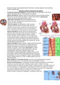
Arterial Thrombosis: Images of inside of heart, died from acute myocardial ischemia.
Blood clot in coronary artery and another. Coronary artery disease. Consequences:
angina, AMI (acute myocardial infarction), death.
Mitral Valve Thrombosis: Mitral valve replacement, like
before, with clots. Consequences are stroke from the clots
going in the brain and death.
Atrial Thrombosis: Atrial fibrillation is where you get to
discordant electrical activity around the atria. They just
fibrillation and don’t pulse, so get blood clots forming on the
inside, so the blood clots break off, go up to aorta and get
strokes, and people die. Consequences are stroke and death.
Arterial Thrombosis: Derived mainly from Arteriosclerosis.
Initiated by fatty streak (gets thicker and thicker) followed by
lipid accumulation, & mature plaque formation on arterial
surface, especially areas of turbulence. The thick fatty streak
can block blood vessels, but get thrombus associated with this,
especially if the layer breaks. Plaques rupture-form haematomas,
exposed surfaces accumulate platelets, form thrombi. So, can get
microthrombi and strokes from that.
Arterial Thrombosis: Arterial occlusion. Platelet and/or fibrin thrombi
detaching from arthro cerotic plaques, and getting stroke and
infarction downstream as a result of that. Downstream tissue
infarction eg Carotid -> stroke; Heart. Risk factors for arterial
thrombosis (atheroclerosis): positive family history, male sex,
hyperlipidaemia, hypertension, diabetes mellitus, gout,
polycythaemia, hyperomocysteinaemia, cigarette smoking, EGG
abnormalities, elevated CRP-IL6-fibrinogen-lipoprotein associated
phospholipase A2, lupus anticoagulant, collagen vascular diseases,
Bechet’s Disease (French c), or CRP, C-reactive protein, EGG, and
electrocardiogram.
Major Categories of Thrombotic Disease: Acute MI, acute peripheral arterial occlusion,
pulmonary embolus, DVT / VTE, ischaemic stroke, atrial fibrillation, COVID.
Venous Thromboembolism: Thrombophilia: laboratory detected predisposition to thrombosis.
Thrombus in one of the deep veins. Hereditary causes: Factor V Leiden 15%, major.
Prothrombin G20210A variant 5% of cases. Protein C deficiency-protein C inactivates factors Va
and VIIIa; deficiency in the degradation of clots because of protein C (or protein S its cofactor)
2%. Protein S deficiency-cofactor for Va and VIIIa inactivation by protein C 2%. Antithrombin
deficiency-inactivates thrombin (IIa) and factor Xa; one of the major
proteins involved in switching off coagulation, so deficiency means will
,carry on and get worse 1%. Consider testing younger patients with unprovoked VTE
especially if family history.
Deep Vein Thrombosis: Venous thrombosis, happens at valves in the leg for
example. Get swirling behind valve. Clot build up-platelet adhesion, Lengthening of
clot or thrombosis. Then a bit flies/falls off. That bit falls off, straight down the vein,
through the heart and into the lungs. Microthrombi or big thrombi blocking blood flow
to a large area of the lung, ending up with profusion defect. Large enough, can kill or
wipe out one lung, maybe both.
Pulmonary Embolism: X Ray. Air space in a lung is red. Right lung, no air space
because of pulmonary embolism of lung. Colour enhanced image of X ray.
Venous Thrombosis: Rudolf Virchow: “Functio Laesa”, Link between cancer &
inflammation (inflammatory cells and cancer). Triad in pathogenesis of
thrombosis: Stasis; Blood components; Vessel Wall. He noted in autopsies clots
in legs and lungs of patients who died of pulmonary embolism (1846). Noticed the link between
leg thrombosis and a lump.
Venous Thrombosis: Virchow’s Triad & Risk Combined risk factors considerably higher
thrombotic risk.
Hereditary and acquired risk factors for venous thrombosis table: Hereditary haemostatic
disorders: Factor V Leiden, Prothrombin G20210A variant, Protein C deficiency, Antithrombin
deficiency, Protein S deficiency, Dysfibrinogenemia, Non-O ABO blood group, DVT in close
relative (especially if unprovoked). Hereditary or acquired haemostatic disorders: raised plasma
levels of factor VIII, fibrinogen, homocysteine. Acquired disorders: Lupus anticoagulant,
Oestrogen therapy (oral contraceptive and HRT), Heparin-induced thrombocytopenia,
Pregnancy and puerperium, Surgery especially abdominal hip and knee surgery, Major trauma,
Malignancy, Acutely ill hospitalized medical patients including cardiac or respiratory failure and
infection and inflammatory bowel disorders, Myeloproliferative disease, Hyperviscosity and
polycythaemia, Stroke, Pelvic obstruction, Nephrotic syndrome, Dehydration, Varicose veins,
Previous superficial vein thrombosis, Age, Obesity, Paroxysmal nocturnal haemoglobinuria,
Behcet’s disease, Diabetes, Immobillity.
Venous Thromboembolism (VTE) Natural History: DVT (deep vein thrombosis) and PE
(pulmonary embolism)-part of the same pathological process. 40% of patients with DVT but no
features of PE clinically have evidence of PE (a small emboli tho) on lung scan. Majority of PE
arise from a leg DVT. Majority of DVT originate in calf venous sinuses. 25% extend proximally to
popliteal vein (the one in the back of the knee) or above. So is in calf, extending up to popliteal
vein or above, you are in trouble. 40% of those (10% of total) embolize. 20% of those (2% of
total) are fatal.
VTE Epidemiology: Overall incidence 1 in 1000 per year (age-dependent). Male (and female),
as increase with age, have increase susceptibility per 100 thousand. Vaccine about thrombosis
and that, this is incidence per 100000 compared to the vaccine which is incidence rates of
hardly anyone per 10 million. PE third most common cause of cardiovascular death. Untreated
mortality of 30%, reduced to 5% with treatment, with anticoagulants especially aspirin and
warfarin. Most common cause of postoperative death and maternal mortality.
Most deaths due to missed diagnosis rather than treatment failure.
,Consequences: pulmonary embolism with DVT, total limb ischaemia, death. Very serious
diseases.
VTE: Post operative risk is high! Hospital Acquired Thrombosis. Up to 50%
of all cases of DVT and PE are acquired in hospital; people are ill, in bed,
not moving around, not moving legs, end up with DVT and PE especially if
they hurt. Post operative Thrombosis, esp postoperative abdominal of hip
surgery (not moving around for sure). Picture-outside GI surgery unit at
hospital, please remember dalteparin fragmin should only be administered
subcutaneously into the stomach or outer aspects of the thigh. So common,
everyone is given this; tells the nurses where to inject them. Very painful,
stings like mad so patients do not like it but must be injected to stop them from getting a DVT.
Venous stasis & immobility: Atrial fibrillation; Congestive HF (heart failure), MI (myocardial
ischemia), varicose veins; Anaesthesia – surgical anaesthesia give them muscle relaxants so
muscles all relax, no tone, pooling within the muscle so start getting thrombi in venous valves;
Long aircraft / vehicle journeys-cheap aircraft, cramped leg space, so get a VT and tunneled off
to hospital; Sedentary lifestyle. Malignancy – TF (tissue factor) / mucin synthesis, so increased
coagulation with them. Inflammation – procoagulant / anticoagulant shift. Shift from
anticoagulant plasma to a procoagulant plasma. IBD (inflammatory bowel disease), SLE,
diabetes, Bechet’s, systemic TB.
Diseases with Elevated D-dimer: D-dimer are the
breakdown products of activated clotting cascade, so the
breakdown products of fibrin. These are all the things where
you get elevated D-dimer. Table: Acute myocardial infarction.
Peripheral arteriopathy. Acute upper gastrointestinal
haemorrhage, other haemorrhage. Aortic
dissection/aneurysm. Acute respiratory distress syndrome.
Arterial or venous thromboembolism. Fibrinolytic therapy.
Atrial fibrillation. Consumptive coagulopathy-DIC. Infection.
Malignancy. Pregnancy. Pre-eclampsia. Sickle cell
disease/haemolysis. Stroke. Superficial thrombophlebitis.
Trauma, burns. Hospitalisation (just sitting around in bed). Old age. Neonatal period. Disability.
How we use D-dimer for measuring coagulations or the state of coagulations within patients.
You have the fibrin being produced through clotting. Factor 8, 8A, fibrin. Plasmin acts on soluble
X-linked fibrin, have our insoluble X-linked fibrin polymer being degraded by plasmin as well.
These are producing D-dimers that we can measure circulating. So we measure D-dimer
release and crosslinked fibers, where the fibrin is being accumulated; end product is D-dimer
release.
Factor V Leiden Protein C Resistance: Part of the prothrombinase complex, factor 5a. Factor
V is a cofactor for factor Xa activation and Thrombin activation;talking about prothrombinase
complex. Protein C cleaves and degrades Factor V. Defect in protein C, cannot cleave factor V,
so prothrombinase complex keeps going on, and have massive coagulation at the end of it. In
Factor V Leiden there is resistance to degradation by Activated Protein C (APC) leading to
enhanced coagulability of plasma. Important female health issue: Pregnancy – 60% of
, thrombosis in pregnancy have Factor V Leiden. 30-40% incidence in venous thrombosis
(VTEs). Oral contraceptive use. Oestrogen Replacement therapy and HRT.
Protein C / S: Can see where APC works. It degrades factor 8. Talking
about the tenase complex (still works), and prothrombinase complex. If
factor 5A is resistant in prothrombinase complex, then this will carry on
going.
Factor V Deficiency: Can get factor V deficiency, but is extremely rare.
Factor V is a cofactor for factor Xa activation and Thrombin activation.
Deficiency results in haemophilia, can be acquired tho. Congenital
deficiency, extremely rare. Variable severity from mild to severe. Acquired:
Liver disease, autoimmunity, Lupus, excessive bleeding.
Hypercoagulability in Pregnancy: Natural adaptive mechanism to limit blood loss post partum,
after labor. Enhanced production of fibrin. Reduced plasmin activity. Raised levels of: thrombin,
fibrinogen (everything that increases coagulability), Plasminogen activator inhibitor -1 (PAI-1),
PAI-2 from placenta. Reduced levels of Protein C.
Management of Thrombotic Complications in COVID19: Major issue now. Consequences of
COVID: microangiopathy in lungs, brain, liver, kidney, there is sepsis-induced coagulopathy
(SIC), and have disseminated intravascular coagulation (DIC). Recent meta analysis of
what happens to patients. Published in 2020. Have number of patients, countries, number
of thrombotic percents, types of events, and mortality! Italy-PE in lung, ischemic stroke is
about arterial thrombosis; myocardial infarction will also lead to ischemic stroke.
France-Circuit clotting-blood support machines like used in the clinic, in surgery; have
clotting within their cardiovascular support circuits. So, clotting is a real problem
with COVID patients.
COVID Associated Coagulopathy: Lower lobe with diffuse alveolar damage with
multinuclear alveolar macrophages (arrows) and striking leukostasis (insert), both
H&E (× 200). Looking at blood vessels, can see some COVID particles. This is in
the endothelium. Virus particles (red arrowhead) are often found in endothelial
cells (white arrowhead, erythrocyte; gray arrowhead, nucleus of endothelial cell);
insert bottom, bar scale 100 nm. Because of the ACE receptor that COVID binds
to, they are expressed highly in lung endothelium. Breathes in virus, replicated in
endothelium like crazy. Damages the endothelium. Endothelium has a profound
anticoagulant effect; destroy the endothelium, then you get coagulation. Patient
3: 72yo male admitted with syncope, fever, cough, and emesis. The past medical history
included coronary artery disease, hypertension, currently untreated polymyalgia rheumatica,
and a history of Merkel cell carcinoma with ongoing local adjuvant radiotherapy (BMI 22.3
kg/m^2). ICU at d4, O2 at d6, died d10 renal and hepatic failure. That is sort of an early lung
disease.
Continued: Inflammation of the capillaries. Neutrophilic capillaritis associated with
microthrombosis (arrowheads) and intra-alveolar hemorrhage. H&E (× 200). The insert shows
endothelial [cell] necrosis with nuclear dust (arrow) and transmigration of neutrophils (× 400).
Patient 1: 78-year-old obese female (BMI 35.2 kg/m2) with hypertension and
an atrioventricular block treated with a permanent dual chamber pacemaker.
The patient died at home, after experiencing a 12-h period of fever with cough


