100% satisfaction guarantee Immediately available after payment Both online and in PDF No strings attached
logo-home
Zeer compacte samenvatting van de anatomie OOG $6.98
Add to cart

Summary

Zeer compacte samenvatting van de anatomie OOG

 4 views  0 purchase
  • Course
  • Institution

Een samenvatting is geen samenvatting meer zodra het oneindig veel pagina's bevat. Met deze documenten zal je de toets zeker halen!! *** TIP: het document afbeeldingen OOG is hierbij erg handig voor algemeen begrip!!

Preview 1 out of 2  pages

  • June 19, 2023
  • 2
  • 2022/2023
  • Summary
avatar-seller
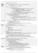
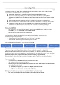
Samenvatting anatomie oog
LAGEN  Sclera
OOGBOL o Harde oogrok. Stevig i.v.m. extra- en intra-oculaire druk. Aanhechting spieren
(BULBUS o Anterieur: cornea
OCULI)  Doorzichtig, geen vaten, zeer gevoelig
 Uvea
o Vaatrijke laag, bestaat uit 3 delen:
 Iris, diafragma rond centrale opening/pupil. Regelt lichtinval in oog d.m.v. spiervezels
 Gekleurd pigment in iris absorbeert licht
 PSZ → m. sphincter pupillae → vernauwen pupil → minder lichtinval
 SZ → m. dilator pupillae → verwijden pupil → meer lichtinval
 Corpus ciliare, anterieure verdikking
 Functie: productie oogkamervocht, lensophanging, accommodatie → m. ciliaris
 Choroidea, vaatvlies → vasculariseert retina en rest van het oog
 Retina (geen contact tussen beide lagen = ablatio retinae, netvliesloslating → blindheid)
1. Pigment laag (buiten), absorbeert licht
2. Neurale laag (binnen, niet anterieur)
 Gele vlek (macula lutea), centraal in retina → zorgt voor scherp zicht
 Fovea centralis: centrale depressie, geen vaten
 “blinde vlek” → plek aanvoer n. opticus en vaten, geen fotoreceptoren (hersenen vullen ‘gat’ in)
 Fotoreceptoren
 Staafjes: 120 mln. Zwart/wit, zeer gevoelig voor licht → nachtzien
 Kegeltjes: 6 mln. → 3 soorten: rood, groen, blauw → kleuren zien, m.n. bij de gele vlek
Compartiment  Voorste segment: bevat oogkamervocht
o Voorste oogkamer: cornea ↔ iris, 3mm
o Achterste oogkamer: iris ↔ lens, 0,06ml
 Achterste segment: bevat glasvochtholte (camera vitrea) met corpus vitreum (glasachtig lichaam)
o Ruimte achter lens, 4,5ml (98% water, 2% collageen)
Oogkamer  Productie door corpus ciliare
vocht  Functie: voeden lens en binnenkant cornea, wordt constant ververst
Verloop:
1. Corpus ciliare
2. Achterste oogkamer
3. Pupil
4. Voorste oogkamer
5. Afvoer via kanaal van Schlemm bij iridocorneale hoek
6. Afvoer via venen
Glaucoom
 Open kamerhoek → afvoer obstructie of overproductie
 Gesloten kamerhoek → obstructie iridocorneale hoek, kan verergerd worden bij verwijden pupil → acuut glaucoom
Lens In een kapsel opgehangen en uitgespannen binnen ring van het corpus ciliare; bol van zichzelf
 Corpus ciliare ontspannen → lens plat, normale dioptrie sterkte
 Corpus ciliare aangespannen → lens ontspannen/bol, hogere sterkte = accommodatie
Functie: licht doorlaten, lichtstralen bundelen, beeld dichtbij scherpstellen door te accommoderen
Cataract (staar): troebeling lens, eiwitten gaan in de loop der jaren samenklonteren → visusdaling, lichtverstrooiing,
blindheid
Visuele baan 1. Uitlopers zenuwcellen retina verlopen naar verzamelpunt in retina; blinde vlek
2. Verlaten daar gezamenlijk het oog als n. opticus
3. Loopt via punt van orbita en canalis opticus de schedelholte in
4. Komen samen bij chiasma opticum
a. Nasale netvlieshelften kruist
b. Temporale netvlieshelften kruizen niet
5. 2 bundels naar visuele schors (occipitale hersenschors)
a. Visuele info van rechter netvlieshelft: linker gezichtsveld naar rechter schors
b. Visuele info van linker netvlieshelft: rechter gezichtsveld naar linker schors
Verdeling bepaald schade aan visuele baan: voor, bij of achter chiasma
Refractie Refractie: lichtbrekend vermogen van cornea en lens, in dioptrieën (D)
afwijkingen o vertepunt: punt verst van oog af gelegen waarbij oog ongecorrigeerd nog scherp kan zien
o nabijheidspunt: punt dichtst bij oog gelegen waarbij oog ongecorrigeerd nog scherp kan zien
 Emmetropie: normaal. Scherpzien zonder accommodatie; vertezien
 Myopie (bijziendheid, -D)
o niet accommodeerbaar, correctie met negatief brillenglas

The benefits of buying summaries with Stuvia:

Guaranteed quality through customer reviews

Guaranteed quality through customer reviews

Stuvia customers have reviewed more than 700,000 summaries. This how you know that you are buying the best documents.

Quick and easy check-out

Quick and easy check-out

You can quickly pay through credit card or Stuvia-credit for the summaries. There is no membership needed.

Focus on what matters

Focus on what matters

Your fellow students write the study notes themselves, which is why the documents are always reliable and up-to-date. This ensures you quickly get to the core!

Frequently asked questions

What do I get when I buy this document?

You get a PDF, available immediately after your purchase. The purchased document is accessible anytime, anywhere and indefinitely through your profile.

Satisfaction guarantee: how does it work?

Our satisfaction guarantee ensures that you always find a study document that suits you well. You fill out a form, and our customer service team takes care of the rest.

Who am I buying these notes from?

Stuvia is a marketplace, so you are not buying this document from us, but from seller OperatieassistentIO. Stuvia facilitates payment to the seller.

Will I be stuck with a subscription?

No, you only buy these notes for $6.98. You're not tied to anything after your purchase.

Can Stuvia be trusted?

4.6 stars on Google & Trustpilot (+1000 reviews)

52510 documents were sold in the last 30 days

Founded in 2010, the go-to place to buy study notes for 14 years now

Start selling
$6.98
  • (0)
Add to cart
Added