100% satisfaction guarantee Immediately available after payment Both online and in PDF No strings attached
logo-home
Summary Systems Neuroscience $18.03
Add to cart

Summary

Summary Systems Neuroscience

 51 views  3 purchases
  • Course
  • Institution

In this summary (based on my own notes and PPT) you can find all the chapters that have been given in the Systems Neuroscience course, except Part II of the Vestibular system. Color code: -Purple= 1. -Dark pink = 1.1. -Light pink = 1.1.1. -Green= 1.1.1.1 - Blue= 1.1.1.1.1 Important abbr...

[Show more]
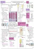
Preview 4 out of 51  pages

  • October 17, 2023
  • 51
  • 2022/2023
  • Summary
avatar-seller
EMBRYOGENESIS SYSTEM NEUROSCIENCE: NERVOUS SYSTEM RETINA
neurulation !!! DEVELOPMENT = invagination of neural tube
= production neural tube, B-vesicles & spinal other factors involved in neural induction:
cord > neuronal plate: NT3
> surface ectoderm
a) dotted line = production notochord > LPM: BMPs
(mesoderm)
→ sends out TF (Shh & Noggin) to
> PM: Twists PATTERNING
> endoderm = molecular & cellular specification area’s
ectoderm
b) ectoderm is going to raise converted to 5 B-vesicles: of developing tissue
c) development butterfly shape > pro divides in 2:
→ from this moment 2 ≠ ectoderms: • telencephalon = cerebral cortex SONIC
> white = surface ectoderm • diencephalon = thalamus, hypothalamus, HEDGEHOG
> blue = neurectoderm (lays pineal gland & retina PROTEIN
elevated on suf. ectoderm) > mesencephalon = midB
> rhom divides in 2: = ventralizing factor prod. by notochord
d) central<<< part butterfly penetrates
• metencephalon → prod. in ventral region: induces
into depth
• myelencephalon floorplate & ventral cell types in
→ formation neuronal pit/groeve
1. surface ectoderm / 2. neuronal plate / neural tube
3. neuronal pit / 4. neuronal crest with cells / DEVELOPMENT VENTRICULAR
5. neuronal tube / 6. spinal ganglion / 7. neuro- SYSTEM
porus cranialis/anterior / 7. neuroporus caudalis/ BMP4
posterior / 9. notochord / 10. primitive knot / = bone morphogenetic protein 4
11. primitive line / 12. somites = dorsalzing factor prod. by notochord
e) indentation (instulpen) neuroectoderm: (sextra info embryology for primitive line etc.) → prod. in dorsal region: induces
2 sides of the pit grow to each other roofplate & commissural intern
& melt together (see as jacket with neuronal tube rise to entire NS & all circuits !!! + shedding part of dorsal cells
2 zippers: begins in the middle & 1 --> move away from tube
zipper closes caudal & other cranial) neuronal tube defects: --> form crest cells: forms
f) creation neuronal tube: completely > anencephaly = error at cranial neuroporus many ≠ cells & tissues
separated from surf. ectoderm → no B bcs of no closure neuronal tube → works together with Sonic P:
(is laying under it) > spina bifida = error caudal neuroporus make a dorsal-ventral axis !!! for
g) between surf. ectoderm & later differentiation
neuroectoderm neuronal crest cells
B-DEVELOPMENT
= special migrating cells neuronal tube forms 3 dilatations pigments cells, cartilage, connective
h) cranial & caudal parts stay open = B vesicles: tissue, N, glia cells,...
temperately > prosencephalon = foreB
= neuroporus cranialis & caudalis > mesencephalon = midB
→ needs to close: o/w neuronal → stays like it is
tube defects > rhombencephalon = hindB

, TbR2 = TF in subventricular zone also tangential migration ~ Tarzan: jump
RHOMBOMERES DEVELOPMENT CEREBRAL from 1 location to another → not
→ radial glia’s -> intermediate progenitors
= regional ≠ in rostro-caudal axis bcs of CORTEX marker completely understand
swelling of the neuronal tube cortex -> ≠ layers → no TbR2?: → eg. intern migration in cortex from
→ only visible at 29day embryo -> less N = B smaller: cortex smaller ventral foreB
→ each rhombomere ≠ size & shape !!! layer = ventricular zone (VZ): contains -> developmental delays
+ express specific Hox genes in N & glia progenitors -> expands massively -> agenesis corpus callosum MECHANISMS THAT
specific combinations (above VZ subventricular zone) CONTROL MIGRATION
→ specific combination will result → first prod. N from preplate (PP) environmental factors also influence on B!!:
> contact-mediated attraction:
in segmental arrangement → their axons + thalamus axons form eg. fetal alcohol syndrome
connection new N~radial glia cell
cranial nerves intermediate zone (IZ) eg. Zika virus: targets progenitor cells
stimulates gap junction formation
→ after this generation N cortical → microencephaly
→ !!! for maturation
Hox genes regulated by morphogens layers II-VI: form cortical layer > contact-mediated inhibition
= signaling factors: directs cell fate & NEURODEVELOPMENTAL
> chemoattraction
tissue development at a distance from split perplate in: DISORDERS > chemorepulsion
their source > marginal zone = future layer I > spina bifida
→ they induce pattern formation > subplate > anencephaly = underdeveloped B MIGRATION DEFECTS
→ response to morphogen altered by > congenital syphilis & toxoplasma
previous patterning tissue these layers form the 6 layers of cortex > schizophrenia not very !!!, but know that it’s possible
> autism → change in cortex factors?
conserved in many spp. > epilepsy -> migration defects
→ same role & orientation in humans & fruit
flies + very homologue APOPTOSIS DIFFERENTIATION
large part of N during development will be in B >100 ≠ N → how so many?
PROLIFERATION apoptosed > genetically programmed fate choices
→ high regulated + key process in develop- > TF in ≠ stages of development
CNS DEVELOPMENT ment (or combo’s of TF)
see B-development RADIAL GLIA > temporal & spatial segregation
long unknown which cell type produced N caspase 9!!! > environmental factors
NEUROGENESIS IN VZ & SVZ → know now it are radial glia cells: → no caspase = bigger B & more B-defects > conserved genetic programming for
located in ventral part & come from neurogenesis
in lining hollow neuronal tube proliferating
cells neuro epithelium cells
MIGRATION > combinatorial transcription code
= neuronal epithelium cells -> divide → divide in 12-24h in other cells: all radial glia cells = substrates for migration (eg. TbR2)
cells neuro epithelium related → produced N keeps hanging on glia cell for → all specify which N it will become
!!! proliferating marker a while
= phospho histone H3 → uses glia as guide to migrate to HUMAN ORGANOIDS
→ ventral part higher proliferation, but final destination = induced pluripotent cells
most of cortex born in dorsal part = radial migration: nicely oriented → used to model human B-disorders
-> 80% N = glutamatergic N & start from ventricular zone & early stages B-development
-> 20% GABAergic N: regulate → eg. cerebral cortex: inside out
excitatory N formed --> 6->5->4->3->2->1

, NEURONAL POLARITY
N reaches final destination
→ start further differentiation: formation
neuronal polarity with input (dendrite)
& output (axons) zones
→ development axonal growth cone

= extension of developing N looking for
synaptic target
→ highly motile, determines growth
direction & interact with ≠ cells

REFINING CONNECTIONS
initial connections not always good: eg. in-
correct target nucleus
→ need regulation
→ neuronal death, axonal retraction
& synapse elimination

SURVIVAL PRESYNAPTIC
CELL
survival regulated by:
> trophic factors prod. by their targets
= neurotrophines: nerve growth factor,
B derived neurotrophic factor &
neurotrophin 3
• secreted P
• NGF !!!!! for survival in development
• all factors promote N extension,
axonal regeneration & synapse
formation
• act via specific R: TRKs

SYNAPSE FORMATION
axonal filaments when connected
properly change in shape
→ forms: presynaptic element,
cleft (15-30nm) & postsynaptic
elements (with R for NT)

, PAVLOVIAN CONDITIONING THE PLASTIC BRAIN
2 !!! concepts for plasticity:
I. MEMORY SIMPLE LEARNED BEHAVIOR IN MEMORY DEFECT AFTER
II. LEARNING = acquisition knowledge/skills APLASIA
through study, experience or being taught HIPPOCAMPAL DAMAGE
→ 1st scientist that looked at learning Eric Kandel: used sea slug to study learning Henri M.: patient with recurrent epileptic
processes in aplasia seizures
& how behavior changes when you learn
A-B) touch siphon -> redraws gill (yellow) → consulted neurologist Scoville: cut piece
= Pavlov
= preventive mechanism temporal lobe away
→ repeat: slug habituates → helped Henri, but Scoville didn’t know
classical conditioning !!!!: dog -> show dog food
-> saliva production -> proceed presentation → now painful stimuli to siphon: function temporal lobe (hippocampus!!)
food with bell -> after while just bell will faster response (dark blue) DISCOVERY SYNAPTIC
induce salivation (light blue = learning period necessary to store new info PLASTICITY IN HIPPO-
→ after learning period bell induces salivation for touch siphon) → Henri couldn’t learn anything CAMPUS CONFIRMS
= Pavlovian conditioning after operation (even family)
HEBBIAN THEORY
HEBBIAN PLASTICITY & LEARNING & MEMORY IN study: isolated hippocampus & made
CONDITIONING ANIMALS sections
90% work biomedical science done in rats & → put stimulation electrode on Schaffer
psychologist Hebb: though synapse collaterals hippocampus &
connections change as result of experience mice
registration electrode CA1 region
→ 2 cells connected via synapse C) neuronal network of aplasia gill-withdrawal → to look at learning Hampton court maze → start stimulation:
→ use of synapse = synapse efficiency↑ reflex: used = labyrinth
→ nowadays not used anymore: now > low freq. stimulation: gives
→ re-suse synapse: changes efficiency > MN = motor N = withdraws gill same response
& builds information > SN = sensory N = register touch gill Morris water maze
> high freq. stimulation: N goes
> 5HT N= serotonine N = register electric back to initial situation, but
classical conditioning according to Hebb: shock → modulates blue connection MORRIS WATER MAZE
after h the response is
US = unconditioned stimulus = food of SN -> MN B) time needed to reach platform intensified = potentiation
CS = conditioned stimulus → see learning curve: after #days
→ learning process results in change in training animals reach platform faster shows that hippocampus has
connections between cells that register C) memory training Hebbian synapse !!!
ringing bell & N that elicit response → remove platform & see in which
(salivation) quadrant animal was most = synapses that get better when used
→ in end ringing bell = salivation → most time in quadrant where (here high freq. stim. potentiates
→ learning = new connections OR platform was originally ↑ synapses-efficiency)
enhancing existing connections D) shows that learning = based on synapses
→ argument that animal was
→ intervention 5HT N modifies connection synapses -> glutamate R: NMDA & AMPA
really learning !!!
SN & MN → NMDA !!! role in potentiation
D) strategies to find platform:
→ learning process situated at level → why NMDA? -> distributed highly
> spatial: uses higher part of B
modulation synapses in cerebral cortex, hippocampus &
> systematic
> repetitive: no use higher parts B striatum

The benefits of buying summaries with Stuvia:

Guaranteed quality through customer reviews

Guaranteed quality through customer reviews

Stuvia customers have reviewed more than 700,000 summaries. This how you know that you are buying the best documents.

Quick and easy check-out

Quick and easy check-out

You can quickly pay through credit card or Stuvia-credit for the summaries. There is no membership needed.

Focus on what matters

Focus on what matters

Your fellow students write the study notes themselves, which is why the documents are always reliable and up-to-date. This ensures you quickly get to the core!

Frequently asked questions

What do I get when I buy this document?

You get a PDF, available immediately after your purchase. The purchased document is accessible anytime, anywhere and indefinitely through your profile.

Satisfaction guarantee: how does it work?

Our satisfaction guarantee ensures that you always find a study document that suits you well. You fill out a form, and our customer service team takes care of the rest.

Who am I buying these notes from?

Stuvia is a marketplace, so you are not buying this document from us, but from seller evevlaemynck. Stuvia facilitates payment to the seller.

Will I be stuck with a subscription?

No, you only buy these notes for $18.03. You're not tied to anything after your purchase.

Can Stuvia be trusted?

4.6 stars on Google & Trustpilot (+1000 reviews)

48298 documents were sold in the last 30 days

Founded in 2010, the go-to place to buy study notes for 15 years now

Start selling
$18.03  3x  sold
  • (0)
Add to cart
Added