Ik behaalde voor dit vak 18/20.
De samenvatting is gebaseerd op de powerpoint en notities van in de les.
11 pagina's
Over de beeldvorming, compartimenten, spieren en lymfestations van de hals
1. Beeldvormingstechnieken
Annotaties die enkel op de illustraties staan => niet te kennen
1 uitzondering: compartimenten --> aangeduid in de presentatie
Annotaties op de radiologische beelden = wel te kennen
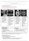
RX Echografie CT MRI
Beperkte waarde: Eenvoudig, snel, Ruim beschikbaar Beste modaliteit voor
klassieke RX vooral beschikbaar Snel beschikbaar beoordeling weke delen
goed vr ossose Geen ioniserende stralen Diepe structuren Geen ioniserende
structuren (botten) => zwangere, kinderen 3D reconstructies stralen: veilig bij
Niet meer gebruikt Enkele voor Ioniserende stralen: zwangere, kinderen
voor beoordeling oppervlakkige structuren enkel aanvragen Nadeel: Beperkte
nek (weke delen) Goed bij stucturen hals indien echt nodig! beschikbaarheid =>
Luchtkolom => allemaal oppervl. , Veel vr onderzoek beperkt aantal
duidelijk zichtbaar geen lucht … hals, vaak bij toestellen
Goede techniek vr 1ste spoedgevallen Duurt langer
evaluatie vd opp. Acute pathologie (onderzoek hals = 30
Halsstructuren (oa min)
schildklier!) Meestal nt als 1ste
diagnose
Computer Tomografie - CT
ZONDER IV-contrast MET IV-contrast
--> Beperkte indicaties --> Standaard
--> gaat voorkeur nt nr uit --> weke delen beter specifiëren: duidelijk contrast
--> bij allergie of slechte nierfct --> bloedvaten intens: hoge densiteit
--> moeilijk onderscheid maken tss bloedvaten, --> anatomische grenzen beter beoordeelbaar
speekselklieren
CT onderzoek vandaag de dag: volummetrisch onderzoek => patient w door CT geschoven => vorming 3D
dataset => reconstructies in verschillende vlakken: axiaal, coronaal, sagittaal
Interventionele beeldvorming
Echografie gebruiken bij procedures
Puncties van afwijkingen in de hals onder echogeleide
FNAC --> fijne naaldaspiratie:
o Met naald in orgaan steken om aantal cellen uit te halen/ fijne biopsie te nemen
o Radioloog in ene hand: echosonde => duidelijk zicht op orgaan & naald
, o In andere hand: spuit met (biopsie)naald => perfect in orgaan terecht komen
o Mogelijk met elk orgaan goed zichtbaar met echografie: in hals:
- Klieren: speekselklieren
- Parotis
- Spieren
- Schildklier ...
Radiologische literatuur beschrijft de anatomie vh hoofd- halsgebied ovv ‘ruimtes’ of ‘loges’ (spaces)
belijnd door fascia
Kennis v deze ruimtes belangrijk:
o Correcte beschrijving aanvragende arts: zelfde taal begrippen
o (Differentiaal) diagnose van afwijkingen hangt af vd stuctuur vd ruimte (verschillende anatomische
structuren in elke ruimte)
- Bv. bij ruimte met vet => gn duidelijke kwaadaardige letsels terug te vinden: weinig tumor
uitgaande v vet
- Ruimte met voornamelijk spierweefsel: tumoren vinden die uitgaan v spierweefsel
- Organen met lymfoiet weefsel => tumoren die daarvan uitgaan
=> zullen bepalen welke aandoeningen in deze ruimtes voorkomen
o Uitbreiding v eventuele tumoren in aanliggende compartimenten bepaalt oncologische stadiëring
De hals is onderverdeeld in
o Suprahyoid en infrahyoid
o Verschillende compartimenten of ruimtes, gescheiden door fasciae
o Onderverdeling zijn arbitrair en kunnen overlappen
Hoe gaat radioloog tewerk?
1. Detectie van een afwijking
2. Bepalen in welke ruimte
3. Normale anatomische structuren van deze ruimte bekijken
4. Beoordelen morfologische kenmerken (Radiologische kenmerken): hoe gedraagt het letsel zich? Hoe
ziet het eruit? Hoe gedraagt het zich nr de naburige organen
& Klinische informatie
5. Diagnose stellen (best guess of DD) => nt altijd mogelijk
➔ Systematische aanpak = belangrijk! + vereist kennis verschillende compartimenten!
Aerodigestieve tractus
Farynx
Nasofarynx (blauw)
o Clivus tot Weke verhemelte
Orofarynx (paars)
o Weke verhemelte tot Valleculae
o Met deeel thv orale caviteit (mond)
Hypofarynx (groen)
o Hyoid tot Cricoid
Larynx
Epiglottis to cricoid (aangeduid met
haakje, geel)
The benefits of buying summaries with Stuvia:
Guaranteed quality through customer reviews
Stuvia customers have reviewed more than 700,000 summaries. This how you know that you are buying the best documents.
Quick and easy check-out
You can quickly pay through credit card or Stuvia-credit for the summaries. There is no membership needed.
Focus on what matters
Your fellow students write the study notes themselves, which is why the documents are always reliable and up-to-date. This ensures you quickly get to the core!
Frequently asked questions
What do I get when I buy this document?
You get a PDF, available immediately after your purchase. The purchased document is accessible anytime, anywhere and indefinitely through your profile.
Satisfaction guarantee: how does it work?
Our satisfaction guarantee ensures that you always find a study document that suits you well. You fill out a form, and our customer service team takes care of the rest.
Who am I buying these notes from?
Stuvia is a marketplace, so you are not buying this document from us, but from seller lorree. Stuvia facilitates payment to the seller.
Will I be stuck with a subscription?
No, you only buy these notes for $5.89. You're not tied to anything after your purchase.