College Research MED-BMS32 Molecular and Cellular Neuroscience (MED-BMS32)
30 views 6 purchases
Course
MED-BMS32 (MEDBMS32)
Institution
Radboud Universiteit Nijmegen (RU)
All the notes from the course MIN32 written in English. Overall, the course MIN32 provides a comprehensive overview of molecular and cellular processes underlying neural processes in the central nervous system in normal conditions as well as in neural disorders. I passed the written exam with an 8....
Molecular and cellular neuroscience lectures
After completion of the course, students are able to
1. Define the major neural cell types of the nervous system
2. Understand the basic mechanism of neuronal communication and plasticity
3. Being able to explain how astrocytes, microglial ad oligodendrocytes affect neuronal communication.
4. Distinguish key molecular signaling pathways critical for learning and memory.
5. Explain how disruptions of molecular pathways lead to impaired cognitive performance in neurodevelopmental disorders.
6. Understand and explain current methodologies to identify and manipulate neural correlates of learning, memory and behaviour.
7. Apply such methodologies for dissecting molecular processes related to specific cognitive tasks in a time and cell-specific way.
8. Use the newly acquired knowledge to critically evaluate and present recent literature reporting new molecular and cellular mechanisms
underlying learning and memory in health and disease
Molecular toolbox for functional analysis of gene mutations underlying cognitive disorders
Lecture 1 – 8th of September 2022
On average, the human brain contains about 100 billion neurons and many more neuroglia which serve to
support and protect the neurons. Each neuron may be connected to up to 10,000 other neurons, passing signals
to each other via as many as 1,000 trillion synapses.
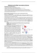
Timing of development of the brain in a mouse: firstly, in E8 to E16, neurons will grow in large amounts. In E14
the astrocytes begin to grow until P13. In E14 also the oligodendrocytes begin to grow but become more
abundant in a later time scale. In the picture you can see this development. Neurons are the output cells and are
supported by the astrocytes and oligodendrocytes. This means that every cell type has a different time of
abundancy in development, this is the same for humans. Non-neuronal cells are typically called glial cells. Rudolf
Virchow searched for connective tissue in the brain. He named this material nervenkitt or neuroglia. There are
different types:
◦ Astrocytes: tripartite synapse gliotransmission (BBB and absorption of neural transmitters)
◦ Microglia: synaptic maturation (development), learning and memory. Remove toxic agents.
◦ Oligodendrocytes: axon myelination
◦ Ependymal cells: ependymal wall of the ventricles and choroid plexus
A presynaptic neuron fires an action potential which goes to the
synapse. The pre synapse and post synapse are very distinct of
each other. The vesicles are in the pre-synaptic side, the post-
synaptic density belongs to the post-synapse. This density is so
dark because there are a lot of receptors and proteins that keep
the receptors in place. The vesicles in the pre-synapse are aligned
with the receptors of the post-synapse. The action potential will
cause the vesicles to go to the membrane, fuse and release their
neurotransmitters in the synaptic cleft to travel to the receptors.
The process of synaptic transmission goes very fast
(milliseconds). The synapse has three parts: pre-synaptic, post-
synaptic side and the synaptic cleft.
Dendritic spines are protrusions of the dendrite where excitatory synapses are formed. Excitatory synapses
(Glutamatergic) are asymmetric. The inhibitory synapses (GABAergic) are very symmetric and are formed on the
dendritic shaft. There is no structure from the dendrite that makes it clear that there is a GABAergic synapse.
One post-synapse can have two PSDs, which means that you have two synapses there. An excitatory synapse
depolarizes the cell with the help of sodium. An excitatory synapse is a synapse in which an action potential in a
presynaptic neuron increases the probability of an action potential occurring in a postsynaptic cell. Positive ions
flow into the cell and the membrane potential will go to -40/-30 mV which will depolarize the cell. 80% of the
synapses in the central nervous system are excitatory (glutamatergic) and present on dendritic spines. Inhibitory
synapses (hyperpolarizing) will have an action potential that will make it more difficult to get an action potential
occurring in a postsynaptic cell.
At rest, a typical neuron has a resting potential (potential across the membrane) -60 to -70 millivolts. This means
that the interior of the cell is negatively charged relative to the outside. Hyperpolarization is when the membrane
potential becomes more negative at a particular spot on the neuron’s membrane, while depolarization is when
the membrane potential becomes less negative (more positive). Depolarization and hyperpolarization occur
when ion channels in the membrane open or close, altering the ability of particular types of ions to enter or exit
the cell. For example:
◦ The opening of channels that let positive ions flow out of the cell (or negative ions flow in) can cause
hyperpolarization. Examples: opening of channels that let K+ out of the cell or Cl- into the cell.
, ◦ The opening of channels that let positive ions flow into the cell can cause depolarization. Example:
Opening of channels that let Na+ into the cell.
The opening and closing of these channels may depend on the binding of signaling molecules such as
neurotransmitters (ligand-gated ion channels), or on the voltage across the membrane (voltage-gated ion
channels). A hyperpolarization or depolarization event may simply produce a graded potential, a smallish change
in the membrane potential that is proportional to the size of the stimulus. As its name suggests, a graded
potential doesn’t come in just one size – instead, it comes in a wide range of slightly different sizes, or gradations.
Thus, if just one or two channels open (due to a small stimulus, such as binding of a few molecules of
neurotransmitter), the graded potential may be small, while if more channels open (due to a larger stimulus), it
may be larger. Graded potentials don’t travel long distances along the neuron’s membrane, but rather, travel
just a short distance and diminish as they spread, eventually disappearing.
Alternatively, a large enough depolarization event, perhaps resulting from multiple depolarizing inputs that
happen at the same time, can lead to the production of an action potential. An action potential, unlike a graded
potential, is an all-or-none event: it may or may not occur, but when it does occur, it will always be of the same
size (is not proportional to the size of the stimulus). To grow synapses you need cytoskeleton (actin in dendritic
spines).
Lecture 2 – 8th of September 2022
Bigger dendritic spines are associated with stronger neural connections. Now underlying mechanisms of this
association are being revealed.
With an action potential, Ca2+ channels will open in the pre-synapse, calcium will flow in which leads to the
transportation of vesicles to the membrane. The vesicles will fuse and let the neurotransmitter go to the post-
synapse. The neurotransmitters will open the receptors here and Na+ will flow in the post-synapse to cause a
new action potential. neurotransmitters can open the ion channels directly or indirectly. The indirect activation
will activate a receptor that will cause a whole cascade that will eventually open the channels.
AMPA receptors are receptors that allow sodium to flow in. NMDA receptors allow sodium and calcium to flow
in. NMDA have very specific properties: they have a magnesium block at resting potential (-60/-70 mV in the
cells) which prevent ions to flow in. Again: the excitatory (glutamatergic) synapses are situated on dendritic
spines. 80% of synapses in central nervous system are excitatory. The dendritic spine with the strongest synapse
is the spin with the largest surface. This is because more receptors can be placed on here, more sodium can go
into the post-synapse, a larger action potential.
There are different distinct phases of synapse development: before and after birth there is a huge phase of
synapse formation. This period lasts until a few years into your childhood. After that people will begin to lose
synapses (synapse elimination) in adolescent years. The system will eliminate the synapses that will not be used.
This is an active phase that is done by microglia. In adulthood you will eventually have synapse maintenance, you
now have the tuned synapse system that will be used in your lifetime. AD will dramatically decrease the amount
of synapses. Developmental disorders will decrease the amount of synapse formation in childhood.
The more AMPA receptors you have, the more depolarization you will have.
The factors that play a role in mediating the action potential in the pre-synapse:
◦ The release probability (the chance that vesicles will fuse and release neurotransmitters when Ca2+
flows in)
◦ The number/size of vesicles
◦ The transmitter content
, Dendritic spines can be coloured by the actin filaments. Actin is only present in the dendritic spines and not in
the dendritic shaft. In the dendrite there is a lot of microtubule, which is not present in the spines. The pathways
that control actin are enriched in the spines. Rho GTPases can transfer extracellular signals to changes in actin,
which will come back in a second. Strong and weak synapses are the basis for learning and memory. Learning
and memory storage in a neuronal circuit is achieved in two ways:
◦ Particular synapses are strengthened or weakened (i.e. in terms of neuronal network theory their
weights are modified), the chance in synapse strength is called synaptic plasticity (a cellular correlate
of learning and memory).
◦ Formation of new and elimination of old synapses rewires and thereby reprograms the circuit.
Long-term potentiation (LTP) and long-term depression (LTD) will change the
strength of the synapse. Both are important for memory and learning. LTP will
strengthen the connection, LTD will weaken it. ‘Cells that fire together wire
together’. Cell A receives input from cell B and C. When Cell A and B are engaged
in the same memory trace, that means that A and B are active at the same time
(B fires at the same time as A). So when they fire together, there will be LTP where
the synapses of both neurons will be strengthened. A and C never fire at the same
time, they can still be connected but they will not function for the same memory.
The synapses between these two are weakened via LTD
(via less AMPAR expression).
LTP: more AMPAR on the surface and a bigger surface of
the post-synapse (dendritic spine needs to grow, thus
actin cytoskeleton needs to grow). How this happens:
Ca2+ will enter the cell and activates CAMKII. This will
activate Rho-GTPases so that the structure will increase.
It will also chance the function (AMPAR will go to the
surface). Example: dentate gyrus has neurons that go to
the CA3, which has neurons that go to CA1. When you
stimulate the CA3 cell, that leads to a response in the
CA1 cell. you can mimic LTP by giving a lot of action
potentials in CA3 (stimuli) for 30 seconds. If you then give another stimulus after that, the response is much
larger. The structure and function has changed so more receptors were available at the grown post-synapse.
Once the action of LTP happened, it can happen for your entire life. The same goes for LTD. The role of AMPA
and NMDA receptors: to get LTP, the post-synapse needs to grow and the amount of AMPAR needs to increase.
Glutamate from the pre-synapse can bind to the AMPAR which will open and Na+ flows in, but this isn’t enough
to depolarize the post-synapse. Glutamate can also bind to the NMDAR but there is still a magnesium block so
nothing happens with the NMDAR. What happens during post-synaptic depolarization, the magnesium block
from the NMDAR will be released, glutamate can bind to NMDAR and Ca2+ will flow into the cell. When both
cells A and B fire together, the glutamate from cell A will go towards the post-synapse of cell B and at the same
time the post-synaptic cell is depolarized. Only in this way (the firing together) the NMDAR will open and a
massive amount of Ca2+ will flow into the cell in a short
time. Ca2+ will activate Calmodulin kinase II (CAMKII)
and protein kinase C which will ensure substrate
phosphorylation via protein kinases. This will ensure
The benefits of buying summaries with Stuvia:
Guaranteed quality through customer reviews
Stuvia customers have reviewed more than 700,000 summaries. This how you know that you are buying the best documents.
Quick and easy check-out
You can quickly pay through credit card or Stuvia-credit for the summaries. There is no membership needed.
Focus on what matters
Your fellow students write the study notes themselves, which is why the documents are always reliable and up-to-date. This ensures you quickly get to the core!
Frequently asked questions
What do I get when I buy this document?
You get a PDF, available immediately after your purchase. The purchased document is accessible anytime, anywhere and indefinitely through your profile.
Satisfaction guarantee: how does it work?
Our satisfaction guarantee ensures that you always find a study document that suits you well. You fill out a form, and our customer service team takes care of the rest.
Who am I buying these notes from?
Stuvia is a marketplace, so you are not buying this document from us, but from seller student300. Stuvia facilitates payment to the seller.
Will I be stuck with a subscription?
No, you only buy these notes for $3.15. You're not tied to anything after your purchase.