100% satisfaction guarantee Immediately available after payment Both online and in PDF No strings attached
logo-home
samenvatting kinesiologie van de heup, KO OL 1ste bach REVAKI $2.68
Add to cart

Summary

samenvatting kinesiologie van de heup, KO OL 1ste bach REVAKI

 11 views  0 purchase
  • Course
  • Institution

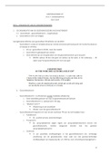
samenvatting ppt +lesnotities kineosiologie van heup, theorie als onderdeel van het vak 'kinesitherapeutisch onderzoek van het onderste lidmaat'

Preview 2 out of 7  pages

  • May 10, 2024
  • 7
  • 2022/2023
  • Summary
avatar-seller
1. Osteo- & arthrologie (herh)...........................................................................................................2
1.1. Rol-glij..........................................................................................................................................2
1.2. Stabiliteit.....................................................................................................................................2
1.3. Mechanische functies..................................................................................................................2
1.4. Heuppathologie...........................................................................................................................2
1.5. Arthrologie...................................................................................................................................2
1.6. Ligamenten..................................................................................................................................2
1.6.1. Capsulaire ligamenten..........................................................................................................2
1.6.2. Intercapsulair ligament........................................................................................................2
1.6.3. Beperkingen vd beweging....................................................................................................2
2. Myologie & beweging (herh).........................................................................................................3
3. Overzicht spieren (herh)................................................................................................................4
4. Kineosiologie.................................................................................................................................5
4.1. Arthrokinematica.........................................................................................................................5
4.1.1. Flexie....................................................................................................................................5
4.1.2. Extensie................................................................................................................................5
4.1.3. Abductie...............................................................................................................................5
4.1.4. Adductie...............................................................................................................................5
4.1.5. Endorotatie...........................................................................................................................6
4.1.6. Exorotatie.............................................................................................................................6
4.2. Stabiliteit heupgewricht...............................................................................................................6
4.2.1. Stabiliteit in frontaal vlak.....................................................................................................6
4.3. Anatomische structuur................................................................................................................6
4.3.1. Inclinatiehoek.......................................................................................................................6
4.3.2. Declinatiehoek......................................................................................................................6
4.3.3. Ligamenten...........................................................................................................................6
5. Dynamisch =/ statisch....................................................................................................................7


Kinesiologie vd heup
1

, 1. Osteo- & arthrologie (herh)
Heup : combinatie v 2 botstukken, 5 ligamenten & 21 spieren
 3D- Beweging : in alle vlakken rond alle assen => kogelgewricht
 Beweging femur tov bekken => stappen (OK)
 Beweging bekken tov femur => voet aan de grond (GK)
 Aantal spieren zorgen vr compessieve krachten & stabiliteit in gewricht
1.1. Rol-glij
 Niet puur rol beweging MAAR naast rollen ook glijden => dankzij kraakbeenlaagje
- Caput femoris : ellipsvormig & volledig bedekt met kraakbeen
- Acetabulum : 3 bekkenbeenderen zorgen elk voor 1/3 vh acetabulum
- Labrum acetabulare : verdiept acetabulum
1.2. Stabiliteit
 Bekomen door gewrichtskapsel & ligamenten (meer dan door omringde spieren)
 Noodzakelijk vr activiteiten (wandelen, staan, lopen)
 Functie ligamenten : zorgt ervoor dat gewricht in bepaalde richting wordt gestuurd &
bewegingen niet mogelijk is
 Ligament scheurt : meer bewegingen mogelijk waar gewricht niet voor gemaakt is
1.3. Mechanische functies
 Ankerpunt vr grote spieren vd romp & onderste lidmaat
 Draagt krachten over vh onderste lidmaat nr bovenste lidmaat
 Brengt gewicht vh bovenlichaam over via tuber ischiadica bij het zitten
 Bekken ondersteunt bekkenbodem (ingewanden)
 Vb : bal gooien in zit-stand => stand : 50% vd kracht komt uit de heup
1.4. Heuppathologie
 3 groepen
 Kinderen (congenitaal)
 Jeugd & sportactieve populatie : overbelastingsletsels
 Ouderen : fracturen & arthrose
1.5. Arthrologie
- Caput femoris : volledig in fossa acetabuli => door labrum acetabuli verdiept
- Gewrichtskapsel
- ligamenten
1.6. Ligamenten
 Geen contractiel weefsel (niet elastisch => om beweging te beperken)
 Taak : beweging af te remmen & gewricht stabiliseren
 4 extra-capsulaire ligamenten= verdikking op kapsel
 1 intra-capsulair ligament
1.6.1. Capsulaire ligamenten
 Ventraal
- Lig. Iliofemorale= lig. Van Bertini
- Lig. Pubofemorale
 Dorsaal
- Lig. Ischiofemorale
 Peri-articulair : zona orbicularis (= ringvormige verdikking vh kapsel)
1.6.2. Intercapsulair ligament
- Lig. Capitis femoris
1.6.3. Beperkingen vd beweging
 Lig. Iliofemorale
- Beperking vd exorotatie in flexie

2

The benefits of buying summaries with Stuvia:

Guaranteed quality through customer reviews

Guaranteed quality through customer reviews

Stuvia customers have reviewed more than 700,000 summaries. This how you know that you are buying the best documents.

Quick and easy check-out

Quick and easy check-out

You can quickly pay through credit card or Stuvia-credit for the summaries. There is no membership needed.

Focus on what matters

Focus on what matters

Your fellow students write the study notes themselves, which is why the documents are always reliable and up-to-date. This ensures you quickly get to the core!

Frequently asked questions

What do I get when I buy this document?

You get a PDF, available immediately after your purchase. The purchased document is accessible anytime, anywhere and indefinitely through your profile.

Satisfaction guarantee: how does it work?

Our satisfaction guarantee ensures that you always find a study document that suits you well. You fill out a form, and our customer service team takes care of the rest.

Who am I buying these notes from?

Stuvia is a marketplace, so you are not buying this document from us, but from seller ellagoosens. Stuvia facilitates payment to the seller.

Will I be stuck with a subscription?

No, you only buy these notes for $2.68. You're not tied to anything after your purchase.

Can Stuvia be trusted?

4.6 stars on Google & Trustpilot (+1000 reviews)

52510 documents were sold in the last 30 days

Founded in 2010, the go-to place to buy study notes for 14 years now

Start selling
$2.68
  • (0)
Add to cart
Added