2.5.4 Innervatie
Hart is bezenuwd door AZS (ortho- en parasympatisch)
Zenuwtakken vormen plexus cardiacus
o Hoog tussen aorta en truncus pulmonalis
o Zakt af naar het hart
o Aan hartbasis nog enkele ganglia
Parasympatische innervatie
o Preganglionaire vezels via n. vagus en rr. Cardiva superiores en inferiors naar plexus
o Vezels linker n. vagus innervatie atrioventriculaire geleiding
o Vezels rechter n. vagus innervatie van sinusknoop
Orthosympatische innervatie
o Komt van bovenste 5 of 6 thoracale spinale sigmenten
o Synaps in sympatische cervicale en hoogthoracale (3 en 4) ganglia
o Postganglionaire vezels via nn. Cardica naar plexus van hart gevoerd
Figuur 2-44: Innervatie van het hart
3 Het vaatstelsel
3.1 Anatomie
Geoxygeneerd bloed wordt door linker ventrikel via aorta in arteries en systeemcirculatie
gepompt
Via rechter atrium en rechter ventrikel wordt gedesocygeneerd bloed via longslagaders naar
longen gepompt komt terug via pulmonaalvenen wordt via linker atrium en linker
ventrikel in aorta gempompt
Indeling in arteries en venen gebeurt op basis van drukverhoudingen en niet op basisi van
niet geoxygeneerd zijn van het bloed
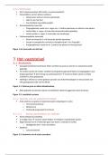
Figuur 3-1: Schema grote en kleine bloedsomloop
Klein gedeelte circulerend volume via lymfatisch stelsel teruggevoerd naar de thorax
Figuur 3-2: Lymfatisch systeem
3.1.1 Arteries van de systeemcirculatie
Al de arteries van grote of systeemcirculatie ontspringen in aorta
o 3cm breed proximaal
o Distaal 2 cm
o Verbreedt progressief in het leven
3.1.1.1 Aorta ascendens en takken
Aorta ontstaat op aortaklep
3 voudige sinus vormen samen bulbus eindigt in sinotubulaire junctie
Begint dorsaal van tr. Pulmonalis gaat naar boven, ventraal en rechtts
o Opstijgende deel = aorta ascendens
Zijtakken:
o Coronairarteriën
Figuur 3-3: Anatomie van de aorta ascendens
,3.1.1.2 Arcus aortae en takken
Begint waar aorta naar dorsaal en links draait en bovenste mediastinum binnendringt
Arcus maakt bocht over linkerhoofdstambronchus over craniale en dorsale zijde
o Kuist trachea en esophagus aan linkerzijde
Vanaf ligamentum arteriosum (hier discrete vernauwing van lumen aortae)
o Aorta loopt voort als aorta descendens
Thoracaal gedeelte: aorta descendens thoracalis
Abdominaal gedeelte: aorta descendens
3 takken ontspringen craniaal op aortaboog van rechts naar links:
o Truncus brachiocephalicus
Rechts naast trachea
Loopt craniaalwaarst naar rechts
Splitst na 5cm in:
A. carotis communis dextra
A. subclavia dextra
o A. carotis communis sinstra
o A. subclavia sinistra
Soms ontspringt nog kleine tak op aortaboog en loopt naar onderpool schildklier
a. thryoidea ima
Figuur 3-4: Arcus aortae
Figuur 3-5: Truncus brachiocephalicus
A. carotis communis
o Loopt stijl craniaalwaarts
o Samen met N. vagus en v. jugularis interna in vagina carotica (bindweefselschede)
Bedekt door m. sternocleidomastoideus
o Splitst aan bovenrand van schildkraakbeen in interna en externa
o Dorsale zijde van splitsing glomus caroticum
Neuro-endocrien orgaan
Werkt voornamelijk als chemoreceptor
o Laatste stuk is gedilateerd (sinus caroticus) hier baroreceptoren gelegen
Figuur 3-6: arteria carotis communis
o A. carotis externa
Verloopt craniaal-lateraalwaarts
Onderkruist achterste buik van m. digastricus en m. stylohyoideus en dtingt
in parotis
Splitst tussen uitwendige gehoorheng en temperomandibulair gewricht in 2
eindtakken:
A. maxillaris
A. temporalis superficialis
Figuur 3-7: Arteria carotis externa
Zijtakken:
A. thyroidea superior
o 1ste ventrale tak
o Buigt naar caudaal naar bovenpool van schildklier
o Geeft a. laryngea superior af
Doorboort samen met n. laryngei superioris de
membrana thyroidea
Verzorgt binnenzijde van de larynx
, A. lingualis
o Beschrijft bocht over os hyoideum
o Loopt ventraalwaarts tussen m. hyoglossus en genioglossus
o Eindigt als a. profunda linguae
A. facialis
o Ontstaat vlak na a. lingualis
o Schuift achter m. digastricus en m. stylohoideus naar gl
submandibularis verwekt hier groeve en bevloeit die
o Voorrand m. masseter doorboort oppervlakkig halsfacia
en draait rond onderrand van mandibula
o Loopt verder langs mondhoek, neusvleugel om medidale
ooghoek te anastomoseren samen met a. dorsalis nasi (tak
van a. ophtalmica
o Verzorgt stuk van farynx, gl. Submandibularis en onderste
deel van gelaat
A. occipitalis
o Ontspringt dorsaal tegenover a. facialis
o Bevloeit achterhoofd
o Veroorzaaakt groeve in proc. Mastoideus
A. auricularis posterior
o Ontspringt dorsaal
o Verloopt in sleuf tussen oorschelp en proc. Mastoideus
A. pharyngea escendens
o Ontspringt procimaal en mediaal op a. carotis externa
o Verloopt lateraal van pharynx naar schedelbasis
Eindtakken:
Figuur 3-8: Eindtakken van arteria carotis externa
A. temporalis superficialis
o Zet richting van carotis externa voort
o Wordt door eerste stuk bedekt met parotis
o Loopt hogerop tussen uitwendige gehoorgang en collum
mandibulae
o Loopt verder naar jukboog hier goed palpabel
o Splitst hogerop in a. temporalis superficialis
R. frontalis
R. parietalis
A. maxillaris
o Ontstpringt in rechte hoek van a. carotis externa binnen
parotis
o Loopt anterieurwaarts tussen caput mandibulae en lig.
Sphenomandibulare
o Dringt tussen 2 koppen van m. pterygoidei
Lateraal of tussen 2 mm.
o Loopt verder naar fossa pterygopalatina
o Eindigt in verschillende takken
van het kaakdeel:
A. auricularis profunda
The benefits of buying summaries with Stuvia:
Guaranteed quality through customer reviews
Stuvia customers have reviewed more than 700,000 summaries. This how you know that you are buying the best documents.
Quick and easy check-out
You can quickly pay through credit card or Stuvia-credit for the summaries. There is no membership needed.
Focus on what matters
Your fellow students write the study notes themselves, which is why the documents are always reliable and up-to-date. This ensures you quickly get to the core!
Frequently asked questions
What do I get when I buy this document?
You get a PDF, available immediately after your purchase. The purchased document is accessible anytime, anywhere and indefinitely through your profile.
Satisfaction guarantee: how does it work?
Our satisfaction guarantee ensures that you always find a study document that suits you well. You fill out a form, and our customer service team takes care of the rest.
Who am I buying these notes from?
Stuvia is a marketplace, so you are not buying this document from us, but from seller klarabukasa. Stuvia facilitates payment to the seller.
Will I be stuck with a subscription?
No, you only buy these notes for $3.95. You're not tied to anything after your purchase.