Summary Keuzevak Infectieziekten pre-master Health Sciences blok 3
8 keer bekeken 0 keer verkocht
Vak
Infectieziekten (AP_470905)
Instelling
Vrije Universiteit Amsterdam (VU)
Boek
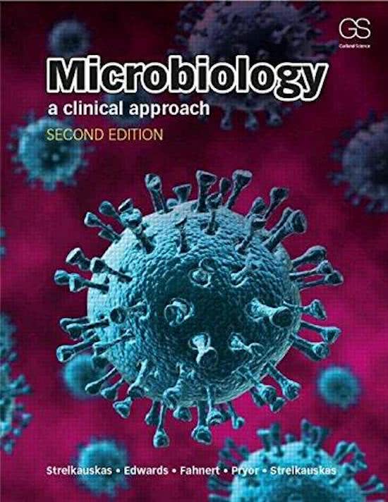
Microbiology
Hierbij de samenvattingen van het keuzevak infectieziekten van de pre-master Health Sciences aan de VU. Met de totale samenvattingen haal je een goed cijfer voor het tentamen infectieziekten.
Keuzevak Infectieziekten pre-master Health Sciences blok 7
Keuzevak Infectieziekten pre-master Health Sciences blok 6
Alles voor dit studieboek
(24)
Geschreven voor
Vrije Universiteit Amsterdam (VU)
Health Sciences pre-master
Infectieziekten (AP_470905)
Alle documenten voor dit vak (7)
Verkoper
Volgen
mirjamdeelen2
Voorbeeld van de inhoud
Chapter 6 (112-127): Transmission of Infection, the Compromised Host,
Epidemiology and Diagnosing Infections
Diagnosing infections
The two key criteria of any diagnostic method are specificity and sensitivity. Specificity
means a test allows us to identify a certain pathogen reliably and correctly. A very sensitive
test permits the detection of very small amounts of the pathogen or antibodies against it.
Growth-based diagnostics
Culturable microorganisms such as bacteria can grow on solid or in liquid media, which
contain all nutrients and other necessary ingredients. Some pathogens are very challenging
to grow, for example legionella pneumophila. Selective media suppress the growth of some
organisms, elective media support the growth of only the organism of interest and
differential media show differences between organisms in mixed samples.
After the mixture of organisms from the sample has been grown in the chosen media, the
suspected pathogen needs to be isolated in pure culture. For cultures containing only the
pathogen, colony morphology can be assessed. The cell morphology is analyzed using a
microscope after the cells have been stained.
Growth-based diagnostic methods can also include analyzing the pathogen’s physiology. A
range of nutrient media is prepared, which are the same except for one component such as
the carbon source. By determining where the pathogen can or cannot grow, we can narrow
down what species it could be. Often there are components added to the test medium that
result in a change of color during fermentation.
Serology-based diagnostics
An alternative is identifying the pathogen based on its antigen, including the production of
antibodies that specifically bind to this antigen. We can either use isolated or manufactured
antibodies to detect pathogen antigens or host antibodies in the sample. Such serological
analysis usually uses blood as the sample, which can be tested directly.
Biotechnology and health
Over time, scientists have developed the ability to use the basic cellular processes in
microbes to create a large number of useful biotechnological tools. Despite the diversity of
cell types in nature, most have the same basic properties: they all need energy to function,
that energy is supplied in chemical form and they all synthesize proteins from a template.
These characteristics provide opportunities for external manipulation.
1. Recombinant DNA technology:
The two-strand structure of DNA has an essential role in the ability of scientists to take apart
a DNA molecule and to recombine it either in different configurations or with DNA from
different organisms. Scientists are able to cut out a piece of DNA (a gene), insert it into DNA
from another organism to make a piece of recombinant DNA. The most commonly used
1
,vector is the plasmid. The recombinant plasmid can then be introduced into a host cell to be
amplified, and sometimes the protein coded for by the original piece of DNA can be made.
Recombinant DNA technology is the foundation of virtually all biotechnological research and
development related to pharmaceutical manufacturing, for example the production of
antibodies for research, therapy and diagnostics.
The standard method for amplifying desired stretches of DNA is the polymerase chain
reaction (PCR), in which a large amount of DNA can be replicated in only a few hours. The
segment of DNA to be amplified is mixed with special short stretches of DNA, the four
nucleotides and an enzyme called DNA polymerase to produce large numbers of copies of
the target DNA.
2. Monoclonal antibodies:
In 1975 Kohler and Milstein developed an advanced type of cell culture for creating
antibodies from a single cell such that all populations of those antibodies are exactly the
same in their specificity and binding abilities. The single cell from which the antibodies are
produced is called a clone cell, so the antibodies are called monoclonal antibodies.
Antibody-producing cells have a relatively short life span. Malignant tumor cell lines are
virtually immortal but incapable of producing antibodies. Fusing them creates a hybridoma, a
hybrid of nuclei from the two cell lines. Hybridomas became the tool for producing
monoclonal antibodies. The production of monoclonal antibodies via hybridomas requires
the use of mice and is prohibited in many countries.
Monoclonal antibodies can be used for everything from diagnostic tests to cancer therapy.
Scientists are able to attach or conjugate toxins and drugs onto these antibodies and then
use them as delivery vehicles. Monoclonal antibodies can be used to attack specific parts of
the immune system of an organ-transplant patient to prevent rejection while leaving the rest
of the immune system free to fight pathogens.
We can produce functional antigen-binding fragments in bacterial systems, but there is even
further use of recombinant escherichia coli in this context. The genetic information for the
antibody component is inserted into the phage genome joined to DNA that codes for a
protein on the outside of the phage, so the protein produced is the antibody fragment joined
to the viral protein. New phages display the antibody fragment to the environment, this is
called phage display.
Usually in phage display we do not start with just the genetic information for one antibody
fragment, but with a library of genetic information. This helps to identify the fragment that
binds best to the antigen. The binding abilities of the displayed antibody fragment can be
improved by repeating selection rounds. After the selection, the genetic information of the
antibody fragments is taken out of the phage vector and inserted into a vector that allows
the production of the antigen fragment only.
2
, The genetic information of the antibody fragments can be genetically fused to marker
proteins such as fluorescent proteins or alkaline phosphates. These markers are the basis of
detecting results in serology-based diagnostics.
Serology-based diagnostic methods
Serology-based diagnosis of infectious diseases is based on an in vitro antigen-antibody
reaction. This reaction is optimal if both antigen and antibody are present at the same
concentration. Samples are usually diluted across a range of concentrations to ensure
successful detection. The lowest concentration of sample at which a reaction can be
detected is called its titer. When antibodies and antigen bind in large amounts, they become
insoluble and therefore visible.
An unknown antigen in the sample can be identified or detected using a known antibody,
which is called the Gruber reaction. The opposite is the Widal reaction, in which a known
antigen is used to spot an unknown antibody in the sample.
Depending on the amount of sample available, a suitable sensitive method is used.
Precipitation detects 0.1-1 mg of antigen, and immunofluorescence 50 ug, making them the
least sensitive. Active agglutination detects about 1.5 ug of antigen, passive agglutination up
to 5x more. Both radioimmunoassay and enzyme immunoassay allow detection of 0.1-1 ng.
1. Precipitation:
Performed in test tubes, a successful reaction can be seen as a white ring (ring test). If we
trap antibodies in a gel matrix and put the antigen sample in a well within that gel, we can
use much smaller amounts. To speed up the movement of proteins in the gel, we can apply
an electric current (electroimmunodiffusion).
2. Agglutination:
Works on a smaller scale and allows faster results by coupling one of the reaction partners to
a bulky component such as erythrocytes or latex beads. Without a reaction the solution
appears milky, when reaction takes place we can observe that the latex beads agglutinate.
3. Immunoblotting:
The immunoblot permits the analysis of a mixture of antigen or antibodies. Proteins are
separated by using an electric current (gel electrophoresis) and then transferred to a
membrane. The membrane is then incubated in a solution containing antibodies. This
procedure is called a Western blot.
The antibodies bind to the antigen on the membrane. The binding is detected via a signal
that can be measured. The antibody can be conjugated to a fluorescent molecule (a
radioisotope), or an enzyme that catalyzes a chemical reaction producing a color change or
light. Alternatively, this antibody is not labeled itself, but a second labeled antibody is used
that binds to the first antibody. We know that the first antibody is from a human, the second
antibody would be from an animal whose immune system has been challenged with human
3
Voordelen van het kopen van samenvattingen bij Stuvia op een rij:
Verzekerd van kwaliteit door reviews
Stuvia-klanten hebben meer dan 700.000 samenvattingen beoordeeld. Zo weet je zeker dat je de beste documenten koopt!
Snel en makkelijk kopen
Je betaalt supersnel en eenmalig met iDeal, creditcard of Stuvia-tegoed voor de samenvatting. Zonder lidmaatschap.
Focus op de essentie
Samenvattingen worden geschreven voor en door anderen. Daarom zijn de samenvattingen altijd betrouwbaar en actueel. Zo kom je snel tot de kern!
Veelgestelde vragen
Wat krijg ik als ik dit document koop?
Je krijgt een PDF, die direct beschikbaar is na je aankoop. Het gekochte document is altijd, overal en oneindig toegankelijk via je profiel.
Tevredenheidsgarantie: hoe werkt dat?
Onze tevredenheidsgarantie zorgt ervoor dat je altijd een studiedocument vindt dat goed bij je past. Je vult een formulier in en onze klantenservice regelt de rest.
Van wie koop ik deze samenvatting?
Stuvia is een marktplaats, je koop dit document dus niet van ons, maar van verkoper mirjamdeelen2. Stuvia faciliteert de betaling aan de verkoper.
Zit ik meteen vast aan een abonnement?
Nee, je koopt alleen deze samenvatting voor $4.84. Je zit daarna nergens aan vast.