Summary Notes about some histology slides viewed in class, with photos of them under the microscope.
3 views 0 purchase
Course
A100 (MEDICINE)
Institution
Medical University Pleven
In this document there are notes took during my histology class, I added then photos and some more informations.
The photos are pretty useful because they are slides viewed under the microscope.
GLANDULA THYROIDEA
Staining: HE
Hematoxylin and eosin is a particular type of staining which
consists in two different types of staining, the hematoxylin
which is a basic staining and the eosin which is an acidic
staining . Basic dyes stains the basophilic tissue component
like the cell nucleus because it contains nucleic acids , but
also it urnderlines membrane proteins. Basophilic tissue
component are those acid parts, so basic dyes have affinity
for acidity.
On the other hand , the eosin is an acidic dyes that stains the
cytoplasm red, and it has affinity for acidophilic tissues.
This duo (hemtoxylin and eosin) Is one of the most common
staining used in histology.
Epithelium: SIMPLE CUBOIDAL EPITHELIUM
In this slide we can recognize a particular type of epithelium which is the simple cuboidal epithelium.
Epithelium covers body surfaces ,Lines body cavities and constitutes glands. Epithelium is an avascular
tissue composed of cells that cover the exterior body surfaces and line internal closed cavities and body
tube. But di epithelium is classified according to two factors :
the number of cell layers and the shape of the Surface cells.
Simple: when it is One cell layer thick
Stratified : when it has two or more cell layers
the individual cells death compose an epithelium are described
as:
squamous: when the width of the cell is greater than its high (
larghezza Maggiore di lunghezza, quasi appiattite).
cuboidal: when width , depth and height are approximately the same
,columnar: when the height is more than the width
in this case the epithelium is simple so there is just one layer of epithelial cells and those cells are cuboidal,
they look like cubes and the nucleus of each cell is large and spherical and is centrally placed. This type of
epithelium can be also resorptive (in general) or secretory, in this case is secretory, and its attached to the
basal membrane , and internally is colloid (non active hormon).
The difference between a follicle and a colloid is that The follicles are lined by follicular cells (simple) that
rest on the basement membrane and have a cavity filled with a homogenous gelatinous material called the
colloid. The collide is composed of thyroglobulin, an iodinated glycoprotein, which is an inactive storage
form of thyroid hormone.
,This epithelium covers the wall of the thyroid gland follicles.The height of the epithelial cells depends on the
phase of normal activity.
The thyroid, or thyroid gland, is an endocrine gland, located in the neck and consists of two connected
lobes. The thyroid gland secretes three hormones: the two thyroid hormones – triiodothyronine (T3) and
thyroxine (T4) – and a peptide hormone, calcitonin.
The thyroid is an endocrine gland and produce the
hormones T3 and T4. It is found in the anterior region of
the neck. Its function is to regulate the body’s energy
metabolism and control the development of the central
nervous system. the thyroid follicles are the structural and
functional units of a thyroid gland. These are spherical,
and the wall is made up of a large number of cuboidal
cells, the follicular cells. These follicular cells are the
derivates of the endoderm and secrete thyroid hormone.
When the hormones are product in the thyroid, they are
subsequently released into the bloodstream. The
secretion of thyroid hormones is controlled by the release
of TSH from the anterior lobe of the pituitary gland into
the bloodstream. If blood levels T3 and T4 are high, TRH is
not produced or released. If blood levels of T3 and T4 are
low, the hypothalamus discharges TRH into the hypothal-
amohypophyseal portal system. Release of TRH stimulates specific cells within the anterior lobe of the
pituitary gland to produce TSH, which in turn
stimulates the thyroid to produce and release more thyroid hormones. As the thyroid hormone levels rise,
the negative feed- back system stops the hypothalamus from discharging TRH. Using the same mechanism
of negative feedback regulation, thyroid hormones also act on the thyrotropes in the anterior lobe of the
pituitary gland to inhibit their secretion of TSH.
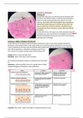
What we can see in the slide:
1. Cuboidal Follicular cells (thyrocytes) -------- simple cuboidal secretory epithelium
2. Parafollicular Cells
3. Sinusoidal Capillaries
4. Colloid (inside thyroid hormone)
Quello cerchiato è un follicolo , all’interno possiamo vedere la sostanza colloide , in un rosa piu scuro , e
attorno le cellule cuboidi del tessuto epitheliale cuboide , e al centro il nucleo sferico.
, SMALL INTESTINE
Staining: HE
Hematoxylin and eosin is a particular type of staining which consists in two
different types of staining, the hematoxylin which is a basic staining and the
eosin which is an acidic staining . Basic dyes stains the basophilic tissue
component like the cell nucleus because it contains nucleic acids , but also it
urnderlines membrane proteins. Basophilic tissue component are those acid
parts, so basic dyes have affinity for acidity.
On the other hand , the eosin is an acidic dyes that stains the cytoplasm red,
and it has affinity for acidophilic tissues.This duo (hemtoxylin and eosin) Is
one of the most common staining used in histology.
Epithelium: SIMPLE COLUMNAR RESORPTIVE EPITHELIUM
Epithelium covers body surfaces ,Lines body cavities and constitutes glands.
Epithelium is an avascular tissue composed of cells that cover the exterior
body surfaces and line internal closed cavities and body tube. But di
epithelium is classified according to two factors : the number of cell layers
and the shape of the Surface cells.
Simple: when it is One cell layer thick
Stratified : when it has two or more cell layers
the individual cells death compose an epithelium are described as:
squamous: when the width of the cell is greater than its high ( larghezza
Maggiore di lunghezza, quasi appiattite)
cuboidal: when width , depth and height are approximately the same
columnar: when the height is more than the width
in this case in the small intestine we can find just one layer so its simple and
then as regars the shape , cells are columnar, and they also have one end
resting on the basal lamina. They have a large elongated nucleus which is
located at the base of the cell, these cells may have micorvilli on their surface
(brushlike or striated border), and sometimes epithelial columnar cells may
be ciliated or non ciliated. An intestinal villus (finger-like projection). Surface
has velvet appearance because of microfolds called villi (microvilli) forming
the brush border. The function is to increase absorption area.
As regards th structure of the small intestine, there are four layers :
The mucosa layer , submucosa layer, muscolaris propria and the serious layer.
-the mucosa layer or tonaca mucosa is the most external One and it can be divided itself in three other
layers like :
the simple columnar epithelium layer,
The benefits of buying summaries with Stuvia:
Guaranteed quality through customer reviews
Stuvia customers have reviewed more than 700,000 summaries. This how you know that you are buying the best documents.
Quick and easy check-out
You can quickly pay through credit card or Stuvia-credit for the summaries. There is no membership needed.
Focus on what matters
Your fellow students write the study notes themselves, which is why the documents are always reliable and up-to-date. This ensures you quickly get to the core!
Frequently asked questions
What do I get when I buy this document?
You get a PDF, available immediately after your purchase. The purchased document is accessible anytime, anywhere and indefinitely through your profile.
Satisfaction guarantee: how does it work?
Our satisfaction guarantee ensures that you always find a study document that suits you well. You fill out a form, and our customer service team takes care of the rest.
Who am I buying these notes from?
Stuvia is a marketplace, so you are not buying this document from us, but from seller federicalolli03. Stuvia facilitates payment to the seller.
Will I be stuck with a subscription?
No, you only buy these notes for $16.88. You're not tied to anything after your purchase.