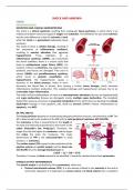
SHOCK AND ANAEMIA
SHOCK
PATHOPHYSIOLOGY
DEFINITION AND CLINICAL MANIFESTATIONS
The shock is a clinical syndrome resulting from inadequate tissue perfusion, in which there is an
imbalance between need and supply of oxygen and substrates. This definition fits also with ischemia,
but the main difference is that the ischemia is local,
while the shock is a generalised inadequate tissue
perfusion.
The result of shock is cellular damage, resulting in
the production of inflammatory mediators,
resulting in vascular alteration that aggravate
hypoperfusion. These mediators (e. g.
inflammatory cytokines, DAMPs, etc.) exacerbate
the shock condition; there is a vicious cycle that
leads to multisystemic organ failure (MOF). For
instance, if there is a blood loss, the organism is less
perfused, and tissues are damaged. They start to
release DAMPs and proinflammatory cytokines,
which result in systemic vasodilation and
hypoperfusion. The hypoperfusion causes a
decrease in the blood pressure, which in turns
decreases the blood flow in tissues, thus causing a further tissue damage, which increases
inflammatory mediator production. This sustained damage and hypoperfusion will give rise to an
irreversible major inflammation.
The initial clinical manifestations of shock are haemodynamic alterations (tissues are hypoperfused)
and organ dysfunction (tissues are damaged), mainly multiple organ dysfunction. The metabolic
dysfunction causes a conversion to anaerobic metabolism at first, which then can develop into cellular
dysfunction (change in ionic gradient, pH), death by necrosis (DAMPs release, inflammation) or
apoptosis, and MOF.
BP, TPR, AND CO
The tissue perfusion depends on maintaining adequate perfusion pressure, and therefore on BP. The
BP is determined by the product of the CO and the total peripheral resistance (BP=COxTPR).
The resistance to flow is proportional to the length of
the tube and inversely proportional to the fourth of the
radius (Poiseuille’s law, R=8L/r4). Therefore, the
longer the tube the higher the resistance, and the larger
the radius the lower the resistance. The major
determinant of TPR is the vasoconstriction in small
vessels (i. e. arterioles).
The cardiac output (CO) is given by the production of the
ejection volume (or systolic output) and the heart rate
(CO=EJxHR) thus the average blood pressure is:
𝒂𝒗𝑩𝑷 = 𝑬𝑱 × 𝑯𝑹 × 𝑻𝑷𝑹
Therefore, if causes of shock wanted to be identified, it would be checked these three parameters.
SYSTOLIC OUTPUT DETERMINANTS
The systolic output is controlled by three parameters, which are:
• Preload (end-diastolic volume, EDV): it is the volume of blood in the ventricle at the end of
ventricular relaxation; it is dependent on venous return and blood volume; this is related to
, the Frank-Sterling law, which states that the heart is a permissive pump, and the stroke
volume is determined by the ventricular end-diastolic volume.
• Afterload (vascular impedance): it refers to the
resistance to flow; it can be increased in case of
hypertension or aortic valve stenosis.
• Myocardial contractility: it refers to the intrinsic
contractile capacity of the heart; it is decreased in case
of infarction, genetic cardiomyopathies, severe
bradycardia, and ventricular fibrillation.
FRANK-STERLING LAW
The Frank-Sterling law, or intrinsic law of the heart states that by increasing the EDV there is a linear
increase of the vascular performance (i. e. SV), up to the maximal capacity. Therefore, the more the
heart is filled, the more blood will be pumped,
and since the heart filling is determined by the
venous return and the blood volume, even the
SV will be affected by it.
When an individual moves from resting condition
to walk, the EDV is not largely changed (even
decreased sometimes), but the SV is increased.
This is caused by the activation of the adrenergic
response, which have inotropic (increase cardiac
contractility) and chronotropic (increased heart
rate) effects. The venous return is always the
same, but the SV is different due to different
blood demand.
However, if the heart is intrinsically diseased, the adrenergic response is always present, but the heart
cannot pump correctly. In the case of lack of cardiac contractile strength, the heart is filled
continuously with blood, which causes an increase in the EDV, but the SV is reduced. This is what is
observed in dilated cardiomyopathies, in which the heart due to different problems (e. g. prior AMI,
cardiomyopathies, fibrillation, etc.) has become dilated, and unable to contract efficiently. This may
cause cardiac insufficiency, congestion, dyspnoea, and pulmonary oedema (increased pulmonary
circulation pressure).
PRELOAD, AFTERLOAD, AND CARDIAC CONTRACTION
The preload is defined as the tension of myofibers before the contraction, that controls the strength
of the following contraction. It depends on:
• Blood volume.
• Venous tone: it determines the amount
of blood that will reach the heart and fill
the chambers.
• Intrathoracic and intrapericardial
pressure: it may prevent the filling of the
chamber with blood (e. g.
hemopericardium,
pneumopericardium).
• Atrial contraction: it contributes in some
extent to the filling of ventricles; the
atrial fibrillation may cause the reduction in preload.
The afterload indicates the resistance to the ventricle opposed by the aorta and the arterial tree, that
the ventricle must win to pump blood in the systemic circulation. It depends on the ventricular and
,aortic pressure gradient, and thus on aortic and systemic blood pressure. A common condition
leading to an increase in afterload is hypertension, as well as aortic stenosis.
The cardiac muscle has an intrinsic contraction property; the coordinated contraction of the
ventricular myocardium determines cardiac output. The contractility is controlled by the preload, the
afterload, and the neurohormonal regulation. This function is impaired in different intrinsic cardiac
conditions, such as hypertrophic cardiomyopathies, arrhythmias, myocardial infarction, and
ventricular fibrillation.
DETERMINANTS DEFINITION FACTORS DISEASES
Blood volume
Hypovolemia
Venous tone
Venous congestion
Tension of myofibres Intrathoracic and
Preload Hemopericardium
before the contraction intrapericardial
Pneumopericardium
pressure
Atrial fibrillation
Atrial contraction
Resistance to the Ventricular-aortic
ventricle opposed by pressure gradient Arterial hypertension
Afterload
the aorta and the Systemic BP Aortic valve stenosis
arterial tress Aortic BP
Hypertrophic
Preload
cardiomyopathies
Myocardial Intrinsic contractile Afterload
Arrhythmias
contractility properties Neurohormonal
Myocardial infarction
regulation
Ventricular fibrillation
COMPENSATION MECHANISMS FOR DECREASE IN BP
SHORT-TERM MECHANISMS
The compensation mechanisms for a decrease in the BP can be performed by different mechanisms.
For instance, after a huge loss of blood (e. g. injury, trauma, etc.) the first immediate response is
neuronal, which is caused by baroreceptors. The
baroreceptors are found in the aortic arch and in the
carotid sinus. The reduced input from arterial
baroreceptors decreases the activity of the vasovagal
centres in the brainstem. The output will be an inhibition
of the parasympathetic cardiac centres and an activation
of the sympathetic cardiac centres, thus resulting in
tachycardia, increased cardiac contractility (chronotropic
and inotropic effect), and arteriolar vasoconstriction. This
condition is called initial shock and occurs in seconds.
The baroreflex will cause vasoconstriction in the areas
that have a reduced metabolic demand (e. g. skin, splanchnic circulation). For that reason, a typical
manifestation of shock is becoming pale, with cold
sweat (adrenergic response) and increased HR.
Moreover, during the rapid neurohormonal response,
the adrenal gland releases epinephrine and cortisol,
which have metabolic effects (glycogenolysis and
gluconeogenesis) and decreased insulin resistance.
Note that the neurohormonal response is fast, but it
cannot last for long time. Indeed, after some seconds
to minutes the problem cannot any longer be
sustained, and other mechanisms should be activated.
, An example of neuronal fatigue is myasthenia gravis. At the beginning the muscle does not have
problems in contraction since neuronal response is present, but after a while problem will arise.
LONG-TERM MECHANISMS
The impossibility to base the compensation only on neuroendocrine regulation causes the necessity
to identify other compensatory, long-term mechanisms. The long-term response to drop in blood
pressure is mediated by the RAAS. The drop
in the blood pressure or the decrease in
sodium concentration in the renal tubule
causes the release of renin by the macula
densa. The renin release in the bloodstream
converts the angiotensinogen into
angiotensin I, which is then converted into
angiotensin II by the angiotensin-
converting enzyme (ACE, mainly in lung
vessels).
The angiotensin II has two effects, the first
is to induce the vasoconstriction (via AT1),
while the second is to induce aldosterone release. The aldosterone release is important to retain
sodium, and therefore also water, thereby increasing blood volume. The combined increase in blood
volume and vasoconstriction increases the systemic BP. Note that angiotensin II can decrease the BP
if it binds to AT2.
Typically, the RAAS activation is accompanied with ADH release. Indeed, increased plasma osmolarity
or reduced blood volume favours ADH release, which will prevent further loss of urinary fluid.
TYPES OF SHOCK
HYPOVOLEMIC SHOCK
The shock is classified into three main types according to the main cause of the drop in the BP, which
are:
• Hypovolemic shock: it is caused by a
decrease in the preload.
• Cardiogenic shock: it is caused by an
insufficiency of the cardiac pump.
• Distributive shock: it is caused by a
reduction in the TPR; differently from
the other two types of shock, paleness
is not present.
The hypovolemic shock is caused by a decreased
in the preload, and it is mainly caused by a
decrease in the blood volume (i. e.
hypovolemia). The true hypovolemia can be caused by two main factors, which are:
• Massive haemorrhage: it can be caused by trauma, GI (e. g. oesophageal varices) or aneurysm
rupture, and internal cavity bleeding; this may be associated with haemorrhagic anaemia,
but the anaemia is observed in chronic conditions.
• Hydrosaline deletion: it can occur during burns, in which the absence of skin does not prevent
water evaporation, and vomiting/diarrhoea, as in the case of secretory diarrhoea (e. g.
cholera, fluid waste, no inflammation).
The preload can be also reduced by other causes, which are not associated to hypovolemia. These
causes are called obstructive extracardiac shock; they are:
Pericardial tamponade: the blood fills the pericardium preventing correct chamber filling.