100% satisfaction guarantee Immediately available after payment Both online and in PDF No strings attached
logo-home
NR507 Final Exam 2024 / NR 507 Week 6 Exam Advanced Pathophysiology Expected Questions and Answers (2024 / 2025) (Verified Answers)- Chamberlain $18.09
Add to cart

Exam (elaborations)

NR507 Final Exam 2024 / NR 507 Week 6 Exam Advanced Pathophysiology Expected Questions and Answers (2024 / 2025) (Verified Answers)- Chamberlain

2 reviews
 32 views  0 purchase
  • Course
  • NR 507 ADVANCED PATHOPHYSIOLOGY
  • Institution
  • NR 507 ADVANCED PATHOPHYSIOLOGY

NR507 Final Exam 2024 / NR 507 Week 8 Exam Advanced Pathophysiology Expected Questions and Answers (2024 / 2025) (Verified Answers)- Chamberlain NR 507 (Latest 2023 / 2024) Final Exam Advanced Pathophysiology - Chamberlain College of Nursing Verified Answers (Graded A+ )

Preview 4 out of 48  pages

  • July 16, 2024
  • 48
  • 2023/2024
  • Exam (elaborations)
  • Questions & answers
  • nr 507
  • NR 507 ADVANCED PATHOPHYSIOLOGY
  • NR 507 ADVANCED PATHOPHYSIOLOGY

2  reviews

review-writer-avatar

By: khadijaabdigaani • 4 months ago

review-writer-avatar

By: Bruce5759wayne • 4 months ago

avatar-seller
APlusAchiever
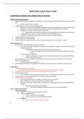
NR507 FINAL EXAM STUDY GUIDE

ALTERATIONS OF RENAL AND URINARY TRACT FUNCTION

URINARY TRACT INFECTIONS (UTI)
 Bacteria from the gut can invade urinary epithelium to cause inflammation and infection anywhere along the urinary
tract.
o Urethra, bladder, ureter, or kidney/s.
 Some individuals are predisposed to developing a UTI.
o more common in women especially when pregnant, sexually active, during post-menopause with estrogen
deficiency, and when being treated with antibiotics where normal bacteria flora is diminished.
o Although less common, men may develop a lower UTI.
 Longer urethra and ureter structures can make it more difficult for bacteria to reach the kidney.
o An indwelling urinary catheter can also contribute to the development of UTI.
o Individuals who experience urinary obstruction, have DM, or neurogenic bladder.
 Can be discussed in terms of severity.
 Can be complicated or uncomplicated.
 Can be discussed according to its location, where it can occur along the urinary tract (lower or upper).

Pathophysiology of UTI
 Bacteria enters and contaminates the lower urinary tract.
o This causes colonization of bacteria in the urethra and the bladder  triggers an inflammatory response in
the lower urinary tract.
 Neutrophils are recruited to the area where the bacteria are present.
 The bacteria multiply which allows them to evade the immune system due to virulent
factors.
o The bacteria can form BIOFILMS.
 A biofilm is any group of microorganisms that allow them to stick to one another and adhere to
surfaces that help them survive.
 If the UTI progresses, or is not treated, or if the patient is immunocompromised, the bacteria can ascend to the
kidneys and colonize there.
o The infection becomes an upper UTI.
 If left untreated, the bacteria can spread into the circulation via the renal veins  bacteremia  septic shock.

UTI Risk factors
 For women, pregnancy is a risk factor.
o Progesterone relaxes smooth muscle that causes stasis of urine  bacteria colonize.
 The female ureter is shorter and allows for entrance of bacteria into the urethra.
 Postmenopausal women.
o Lack of estrogen results in vaginal and urethral dryness  promotes an environment for bacteria to grow.
 Sexual intercourse  can easily introduce bacteria into the urethra; spermicides.
 Indwelling urinary catheterization
o The bacteria will colonize in the bladder and initiate an immune response.
o The neutrophils enter the area to further promote inflammation.
o Fibrinogen accumulates on the catheter  attachment of uropathogens that express fibrinogen-binding
proteins.
 The bacteria will multiply to form biofilms.
 this results in epithelial damage to the urinary tract  kidney infection.
Lower vs. Upper Tract Disorders
 Lower tract disorders
o Urethritis  infection at the opening of the urethra.
o Cystitis  bladder infection
 Can occur in both males and females.
 In males, cystitis may be associated with prostatitis.
o Symptoms:
 Urgency associated with burning on urination.


1

,  Frequency, dysuria, suprapubic pain.
 The urine may appear cloudy and have an odor.
 Upper Tract Disorders
o When the bacteria ascends to the kidney/s  PYELONEPHRITIS.
 Microorganisms associated include: E. coli and Pseudomonas
 Escherichia coli (E. Coli)
o The most common organism contained in the fecal matter that is easily
accessible from the anus to the urethra.
 Staphylococcus saprophyticus, Proteus mirabilis, Klebsiella
 More commonly associated with infections after urethral instrumentation or urinary
tract surgery.
 Also split urea into ammonia  urine alkaline  increases risk of stone formation.
 Dissemination also may occur by way of the bloodstream, and both kidneys are usually involved.
 The inflammatory process is usually focal and irregular, primarily affecting the pelvis, calyces, and
medulla.
 The infection causes medullary infiltration of neutrophils with tubulointerstitial inflammation,
renal edema, and purulent urine.
 In severe infections, localized abscesses may form in the medulla and extend to the
cortex.
 Primarily affected are the renal tubules; the glomeruli are usually spared.
 Rarely but possibly cause acute renal failure.
o Signs and symptoms of Pyelonephritis
 The onset of symptoms is usually acute, with fever, chills, and flank or groin pain.
 Symptoms characteristic of a UTI, including frequency, dysuria, and costovertebral tenderness.
 Older adults may have nonspecific symptoms, such as low-grade fever and malaise.
 There can also be signs of shock if the infection has entered the circulation from the kidney via the
renal vein.
 Classic triad of symptoms: vomiting, flank pain, and fever.
 The presence of systemic signs such as high fever, chills, and tachycardia may suggest severe
infection.

Urine Sample to Determine Lower vs. Upper UTI
 Urine Dipstick
o (+) leukocyte esterase, (+) nitrites
 Leukocyte esterase is an enzyme that is released by the WBCs.
 Qualitative measure of WBCs in the urinary tract.
 Presence of nitrites is highly specific for bacterial infection.
o Can be performed to identify hematuria, proteinuria, and presence of nitrites.
 On microscopic exam of the urine:
o WBC > 5000 hpf and hematuria = Cystitis.
o WBC casts = Pyelonephritis
 Presence of CASTS in the urine  the protein in the lumen of the kidney tubules has solidified,
especially in the nephron.

Uncomplicated Vs. Complicated UTIs
Uncomplicated
 Urinary tract and renal function is normal.
 Individuals are treated for UTI only when they are symptomatic.
o Antibiotics only if patient is c/o symptoms.
 Uncomplicated + symptomatic UTI (cystitis) = 3-7 days course of appropriate antibiotic therapy.
Complicated
 Decreased renal function and abnormal urinary tract.
 Presence of WBC casts = presence of kidney involvement  more complicated treatment plan.
 At higher risk for extensive and permanent kidney damage, as well as sepsis.
o Blood culture may be drawn to identify the causative organism.
 More intervention required.
 Will require IV antibiotics until the patient is afebrile, followed up by a course of PO antibiotics.


2

,  Referral to a urologist if does not respond to antibiotic therapy, or there are recurrent UTIs (3 or more in 1 year), or
presence of hematuria.
o May be a presence of significant renal disease.
 UTI in pregnancy due to ureteral dilation that occurs that increases the risk for pyelonephritis.
o Treatment initiated to prevent damage to the fetus in utero.
 Associated with
o Indwelling catheter
o Renal calculi
o Diabetes
o Pregnancy

Urinalysis to Diagnose a UTI
 Dipstick can be used to identify leukocyte esterase and nitrites.
o Nitrites
 Detect the presence of Enterobacteriaceae (gram negative bacteria) family that converts nitrates
to nitrites.
 Some bacteria are unable to produce nitrites (Enterococcus).
 The presence of nitrites is the most specific finding and has the highest positive predictive value.
o Leukocyte esterase, WBCs and even bacteria  not specific  do not necessarily indicate infection.
 Presence of symptoms + positive urine culture
 Urine examined under microscopy:
o RBCs  > 3RBCs/hpf = abnormal; abnormal morphology of the RBC strongly suggests glomerular disease;
RBCs are often present with hematuria.
o WBCs  > 5 WBCs/hpf = abnormal
o Bacteria  present
o Crystals
o Casts

Patient Education
 Drink more water.
 Although there are differences of opinions, cranberry juice and vitamin C can help to acidify the urine.
 Urinate before and after sexual intercourse to remove bacteria from the urethral area.
 Encourage the female to avoid holding urine for extended periods of time.
 Avoid the use of hygiene sprays and spermicides because they alter the normal microbial flora to enhance the risk for
infection.
 Encourage the female to wipe from front to back after a BM to avoid spreading bacteria to the urethra.
 Encourage showers rather than bathing to avoid the spread of bacteria.

RENAL CALCULI
Pathophysiology of Renal Calculi
 Renal calculi or kidney stones, can be found in the ureter or bladder.
 Urine
o Consists of water and particles (Na+ and K+).
o The particles in the urine combine to form a stone.
 Normally there should be no stone formation in the urine.
o Has inhibitors to prevent stone formation (citrate and magnesium).
o Contains calcium and oxalate.
 Calcium + oxalate = stone.
 Citrate and magnesium will prevent stone formation.
 Individuals get kidney stones due to lack of water ingestion or by increase of particles or solute in the urine or by
decreased inhibitors present in the urine.
 Stone can be easily eliminated in the urine, or if large, can lodge in the ureter.
o Stones that are < 0.5 cm can pass through the urine to be eliminated without difficulty.
o Stones that are > 1 cm are likely to cause an obstruction.
 Most common sites of obstruction:
o Ureteropelvic junction
o Intersection of the ureter and iliac vessels
o Ureterovesicular junction

3

,  Supersaturation: urine becomes oversaturated with certain substances like calcium.
 Nucleation: crystals act as nucleation sites where further crystal deposition can occur.
 Crystal retention: urinary stasis or inadequate urine flow allows crystals to remain in the urinary tract.
 Stone growth and composition: over time, crystals accumulate and grow into stones.

Types of Kidney Stones
 Calcium stone
o Most common type
o Calcium + oxalate, or calcium + phosphate
o Radio dense  can be seen in x-ray.
o Mostly idiopathic in cause
o The individual becomes either hypercalcemic or presents with excess calcium in the urine (hypercalciuria) 
solutes to increase and form a stone.
o Treatment involves prescribing a thiazide diuretic to excrete urinary calcium.
 Struvite stone
o Occurs due to a UTI, most often by proteus, klebsiella, and serratia and Enterobacter species.
o Ammonium + magnesium + phosphate
 The bacteria contribute to the stone formation through the production of enzyme, urease.
 Urea, in the presence of urease converts to ammonia and a byproduct of CO2.
o Makes the urine alkaline which favors stone formation.
o Aka Staghorn stone
o Obstructs the renal calyx.
o Contains irregular, horn-like structures – like a stag’s horn.
 Uric Acid stone
o Found in patient with gout.
o Increase in uric acid.
 Individuals who are at risk for getting gout include those with leukemia and myeloproliferative
disorder; those undergoing chemotherapy.
 Chemotherapy destroys cancer cells  DNA cells contain purine  when broken down,
purine will increase uric acid levels  uric acid stone formation.
o Stones are radiolucent  can not be seen on x-ray.
o Treatment includes hydration and increase alkaline of the urine by giving potassium bicarbonate.
o Allopurinol – anti-gout medication.
 Cystine stone
o Rare type of stone found mostly in children.
o Caused by genetic renal tubule defect  prevents the amino acid, cystine, from being reabsorbed 
formation of cystine stone.
o Can also form staghorn-shaped stones.

Clinical Manifestations of Renal Calculi
 Renal Colic
o Flank or costovertebral angle (CVA) pain.
 Caused by passing of the stone through the ureter with obstruction and spasm.
o The characteristic of pain begins mild then greatly increases causing great discomfort.
o The pain begins in the flank and radiates to the groin.
 As the stone moves, the pain will be in the location where the stone is.
 Hematuria
o Found in 90% of individuals who have kidney stones.
o While passing through the urinary tract, the stone will injure the urinary structures.
o Can be associated with nausea and vomiting.

Diagnosis and Treatment of Renal Calculi
 Diagnosis is confirmed through urinalysis.
o Can have microscopic or gross hematuria.
o The pH of the urine will be identified to help determine the type of stone.
 If it passes, the stone should be taken for analysis to determine what type of stone.
 X-ray of the kidney, ureter, and bladder (KUB) will be performed.


4

The benefits of buying summaries with Stuvia:

Guaranteed quality through customer reviews

Guaranteed quality through customer reviews

Stuvia customers have reviewed more than 700,000 summaries. This how you know that you are buying the best documents.

Quick and easy check-out

Quick and easy check-out

You can quickly pay through credit card or Stuvia-credit for the summaries. There is no membership needed.

Focus on what matters

Focus on what matters

Your fellow students write the study notes themselves, which is why the documents are always reliable and up-to-date. This ensures you quickly get to the core!

Frequently asked questions

What do I get when I buy this document?

You get a PDF, available immediately after your purchase. The purchased document is accessible anytime, anywhere and indefinitely through your profile.

Satisfaction guarantee: how does it work?

Our satisfaction guarantee ensures that you always find a study document that suits you well. You fill out a form, and our customer service team takes care of the rest.

Who am I buying these notes from?

Stuvia is a marketplace, so you are not buying this document from us, but from seller APlusAchiever. Stuvia facilitates payment to the seller.

Will I be stuck with a subscription?

No, you only buy these notes for $18.09. You're not tied to anything after your purchase.

Can Stuvia be trusted?

4.6 stars on Google & Trustpilot (+1000 reviews)

52510 documents were sold in the last 30 days

Founded in 2010, the go-to place to buy study notes for 14 years now

Start selling
$18.09
  • (2)
Add to cart
Added