HAP-20306
Cell metabolism
Energy is stored in the form of ATP (adenosine triphosphate). It is formed by ADP and a free
phosphate, by a reaction called both condensation and phosphorylation (either substrate-level
phosphorylation or oxidative phosphorylation). The break-down of ATP is called hydrolysis.
Glucose oxidation: C6H12O6 + 6 O2 → 6 CO2 + 6 H2O + energy. Figure 1
This has 3 stages: Figure 2
1. Glycolysis (cytosol): glucose → 2 pyruvate (2 ATP & 2 NADH).
2. Krebs cycle (mitochondrial matrix): 2 pyruvate → 2 acetyl CoA (2 NADH & 2 CO2) (linking step);
2 acetyl CoA → oxaloacetate (2 ATP, 6 NADH, 2 FADH2 & 4 CO2).
3. Oxidative phosphorylation (inner mitochondrial membrane): NADH & FADH2 are used in the
electron transport chain which releases energy (O2 needed) → hydrogen ions cross inner
membrane and cause gradient → ATP synthase synthesises ATP by flow of hydrogen with its
gradient (per NADH 2.5 ATP, per FADH2 1.5 ATP). Total ATP production: 32.
If O2 concentration falls: anaerobic conditions. Krebs cycle and oxidative phosphorylation inactive →
pyruvate and NADH accumulate → shutdown glycolysis. But, most cells contain lactate dehydrogenase
(LDH), which converts pyruvate into lactate. NADH can unload an electron with this reaction, and 2
ATP can be produced. Figure 3 This is a dead end: lactate accumulates (acidification of blood and
tissues). Once oxygen is available again: linking step takes place, thus pyruvate converted into acetyl
CoA. Because less pyruvate, LDH reaction turns around and lactate will be converted into pyruvate
(and NADH). This is the Cori cycle.
When there is plenty glucose, it can be transformed into fats, proteins and storage molecules:
glycogen. This reaction, called glycogenesis: happens in the liver, but can also happen in kidneys and
muscle. When glycogen is broken down again into glucose, it is called glycogenolysis. When glucose
concentration in body decreases, a process called gluconeogenesis can take place in the liver: reverse
glycolysis. Figure 4 Glucose can then be formed out of glycerol, lactate and amino acids. This is
important because the central nervous system relies on glucose as energy source.
Fats are primarily stored in adipose tissue. Lipolysis is the separation of glycerol and the fatty acids.
Glycerol can enter the glycolysis pathway, fatty acids can be converted into acetyl CoA by beta
oxidation. If the triglycerides are broken down at higher rate than demand, ketones can be generated.
These can be used as an alternative for glucose by the central nervous system. Fats can also be
synthesised again by a process called lipogenesis.
Proteins can be broken down into amino acids, a process called proteolysis. They are then deaminated
and the remainder is called keto acid (pyruvate or acetyl CoA). These reactions are reversible. Figure 5
Feeding in different animals
One way of feeding is suspension feeding: ciliated surfaces produce currents to draw drifting food in
water into their mouths, or they have a mucous sheet or perform other sweeping movements. One
way of suspension feeding is filter-feeding, filtering food from water that passes through the animals.
,Another type of feeding is deposit feeding. To feed on solid structures, adaptations had to be made:
teeth like structures or teeth are formed, jaw and stomach are adjusted to infrequent meals, but true
mastication is restricted to mammals. Mammals are heterodont, which means that the teeth are
differentiated (homodent means that they are not, first synapsids and crocodiles). They have 4 types
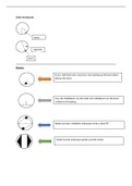
of teeth: Figure 6
1. Incisors (I): simple crown and sharp edges to snip;
2. Canines (C): long, conical crowns to pierce;
3. & 4. Premolars (P) and molars (M): compressed crown with cusps to shear, slice, crush of grind.
The ancestral formula is I 3/3, C 1/1, P 4/4, M 3/3 = 44. Most mammals have two sets of teeth: a
temporary, deciduous, set and a permanent set (exception: molars).
Animals eat more that they gain: prey biomass exceeds predator biomass → big animals eat low in
food chain to minimize energy loss. Smaller animals have a higher metabolic rate, thus eat more
relative to body mass. Animals can evolve into generalists (eat anything) or specialists (eat specifically).
Optimized foraging behaviour is present to challenge the energy gain and cost trade-off. There is for
example a maximal energy gain for the handling time: bird searches black or white worms (hard to
find, more energy gain).
Usefulness = Eblack/Thandling, black & Ewhite/Thandling, white.
Search time = Tsearch, black & Tsearch, white.
Bird continues searching for white after found black: usefulness = Ewhite/(Tsearch, white + Thandling, white)
Eblack/Thandling, black > Ewhite/(Tsearch, white + Thandling, white) → bird eats black worm.
Unicellular eukaryotes and sponges have complete intercellular digestion, using phagocytosis of small
particles (need necessary enzymes and must be capable of absorbing the products). These limitations
were resolved by forming an alimentary system with extracellular digestion. Then food is moved
through the tract by cilia and/or musculature. A true coelom is lined entirely by mesoderm with
muscles on both sides: now intestinal peristalsis is possible, animals have sphincters and independent
movement is possible.
Mammals can be divided into 4 groups:
1. Insectivorous: eat insects, sometimes no teeth, small animals, short intestine, no cecum.
Figure 7
2. Herbivorous: eat grasses (vegetation); browsers/grazers and gnawers. Canines absent or
reduced, fermentation in colon and cecum, many nutrients lost in faeces: eat own faeces
(coprophagy). Ruminanats have four-chambered stomach: rumen (microorganisms),
reticulum, omasum (absorption), abomasum (real stomach) and then small intestine (normal
digestion). Figure 8 & 9
3. Carnivorous: eat mainly vertebrates, large canines, short digestive tract, no or small cecum.
Figure 10
4. Omnivorous: eat plant and animal, cecum mostly poorly developed.
Gastrointestinal system
The gastrointestinal system can be divided into the gastrointestinal tract (GI tract) (where digestion
takes place) and the accessory glands (secreting fluids and enzymes aiding digestion). The wall of the
GI tract has 4 distinct layers: Figure 11
, 1. Mucosa: 3 layers:
- (innermost) mucous membrane: epithelial cells (enterocytes) forming a barrier. Some are
absorptive, some are exocrine (like goblet cells: secrete mucus that protects lining of GI
tract), and some are endocrine.
- (middle) lamina propria: connective tissue (with small blood vessels, nerves, and
lymphatic vessels).
- (outermost) muscularis mucosae.
2. Submucosa: connective tissue with larger blood vessels and lymphatic vessels. Contains
submucosal plexus that is connected with myenteric plexus in muscularis externa (all nerve
cells) → together: enteric nervous system.
3. Muscularis externa: inner circular and outer longitudinal muscle layers.
4. Serosa: fibrous connective tissue and mesothelium (epithelium), which is continuous with the
mesenteries and eventually with the peritoneum.
MOUTH, PHARYNX & ESOPHAGUS
The mouth has salivary glands that produce saliva (all animals except suspension feeders), containing
amylase. Food and saliva mixture then enters pharynx, the upper esophageal sphincter and then
esophagus, whose wall is thin and stretches easily. The upper region has skeletal muscle, the bottom
region has smooth muscle. At the end, there is a lower esophageal sphincter, preventing acidic fluids
from entering the esophagus. Many invertebrates and birds have a crop to store food. In ruminants, 3
of the 4 stomach lobes are found in the esophagus.
By input of the parasympathetic nervous system, secretion of watery saliva increases. By input of the
sympathetic nervous system, secretion of protein-rich, viscous saliva increases.
STOMACH
The stomach of carnivorous and omnivorous vertebrates has 3 regions: fundus, body and antrum.
Figure 12 & 13 The wall of the fundus is thin and can expand easily, the mucosa of the body folds into
rugae, and the antrum has a thick muscle layer for contraction. Fundus and body have gastric pits with
gastric glands that produce and secrete gastric juice. Because of movement, food and gastric juice are
mixed, producing chyme. When the contractions are strong enough, chyme passes the pylorus
sphincter and the pylorus to enter the duodenum. Terrestrial worms and birds can swallow stones and
grit to aid the gizzard for digestion. Rennin is a milk-curling enzyme found in the stomach of ruminants.
To increase the surface area, terrestrial worms have a typhlosole over the full length of the intestine.
Gastric pits contain secretory cells. In the neck, you find neck cells, that secrete mucus. They form the
gastric mucosal barrier because of the acidic environment. Deeper, you find the gastric glands: chief
cells, secreting pepsinogen (inactive form of pepsin), parietal cells, secreting HCl and intrinsic factor,
and G cells, secreting the hormone gastrin (endocrine). Because of the HCl, the stomach is acidic: CO2
+ H2O →(carbon anhydrase)→ H2CO3 → H+ + HCO3-. When HCO3- leaves the parietal cell, Cl- enters the
cell, and eventually leaves the cell again (into gastric lumen). H+ is pumped out of the cell (into gastric
lumen), using ATP. Figure 14 The acid helps converting pepsinogen into pepsin, denatures proteins and
kills bacteria. Acid secretion (and pepsinogen secretion) is stimulated by the parasympathetic system,
gastrin and histamine.
SMALL INTESTINE
The small intestine contains 3 regions: duodenum, jejenum, and ileum. Figure 15 In this part, most
things are absorbed. In the duodenum, pancreatic juice with digestive enzymes and bicarbonate

