100% satisfaction guarantee Immediately available after payment Both online and in PDF No strings attached
logo-home
Ultimate Chapter-to-Chapter CSCS Summary Outline $39.99   Add to cart

Summary

Ultimate Chapter-to-Chapter CSCS Summary Outline

 69 views  2 purchases
  • Course
  • Institution
  • Book

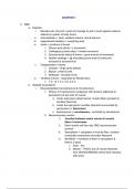
Here's an outlined study guide of all 24 chapters of the book "Essentials of Strength Training and Conditioning - 4th edition" that's commonly used to study for the CSCS Exam by NSCA. This preparatory material will be helpful in understanding the most important big picture concepts in each chapter...

[Show more]

Preview 4 out of 83  pages

  • Yes
  • August 11, 2024
  • 83
  • 2024/2025
  • Summary
avatar-seller
CHAPTER 1

1. MSK
a. Skeleton
i. Muscles can only pull x push but manage to pull or push against external
objects by system of body levers
ii. Axial skeleton = skull, vertebral column, ribs & sternum
iii. Appendicular skeleton = everything else
iv. Joints = junctions of bones
1. Fibrous joint (skull) = x movement
2. Cartilaginous joints (disc) = limited movement
3. Synovial joints (elbow & knee) = good amount of movement
4. Hyaline cartilage = @ articulating bone ends & entire joint
enclosed in synovial fluid
v. Categorization of joints
1. Uniaxial - hinge joints (elbow)
2. Biaxial - ankle & wrist
3. Multiaxial - shoulder & hip
vi. Vertebral column - separated by flexible discs
1. 7 C; 12 T; 5 L; 5 S; 3-5 C
b. Skeletal musculature
i. Musculoskeletal macrostructure & microstructure
1. Fibrous CT (epimysium) contiguous with tendons (attached to
periosteum) at the ends of muscle
a. Under epimysium (deep fascia), muscle fibers grouped in
bundles (fasciculi)
b. Under the epimysium: bundles (fasciculi) surrounded by
perimysium (= fasciculus)
c. Endomysium (= sarcolemma) - encircled by sarcolemma
2. Neuromuscular junction
i. Junction between motor neuron & muscle
fibers it innervates
ii. Each muscle cell has only ONE neuromuscular
junction
iii. Sarcoplasm = cytoplasm of muscle fiber; contains
contractile components of protein filaments
iv. Myofibrils = hundreds of them in sarcoplasm &
there’s 2 types
1. Actin - thin
2. Myosin - THICK; pair of myosin filaments
from CROSS BRIDGE (which then interacts
with actin)

, a. Sarcomere = smallest contractile
unit made up of actin & myosin AND
Within the sarcomere:
i. A-band = alignment of
myosin filaments
ii. I-band: areas in 2 adjacent
sarcomeres that contain only
actin filaments -> decreases
as muscle contracts
iii. Z-line: middle of I band
iv. H-zone: center of sarcomere
(ONLY MYOSIN) ->
decreases as muscle
contracts
v. Sarcoplasmic reticulum (SR) = surrounds each
myofibril
1. Calcium ions stored in the vesicles
2. T-tubules run perpendicular to SR &
terminate between 2 vesicles
3. AP arrives simultaneously thanks to
T-tubules between outlying myofibrils
3. Tendon = attached to bone periosteum
4. Epi -> peri -> endomysium
ii. Sliding-filament theory of muscular contraction = actin filaments at
the end of sarcomere slide in on myosin filaments (pulling z-lines toward
the center of sarcomere); therefore, both H-zone & I-band shrinks ->
rapid, repeated flexion must occur in many cross bridges
1. Resting phase - little calcium present in myofibril (very few myosin
crossbridges bind to actin)
2. Excitation-contraction coupling phase
a. Before myosin cross bridges can flex, they must first attach
to actin filament
b. When SR is stimulated to release calcium ion, calcium
binds with troponin (protein situated at regular intervals
along actin filament)
i. This causes tropomyosin movement & myosin
crossbridges now attach much more rapidly to actin
filament
c. Amount of force produced = number of myosin cross
bridges bound to actin filaments cross sectionally
3. Contraction phase
a. Energy for pulling action = power stroke comes from
hydrolysis of ATP to ADP & phosphate

, b. Another molecule of ATP must replace the ADP on myosin
cross bridge globular head in order for head to detach from
active actin site
i. This allows contraction process to continue or
relaxation process to occur
4. Recharge phase
a. Contraction can occur over and over again as long as
calcium available in myofibril & ATP available to
uncouple myosin from actin
5. Relaxation phase - relaxation occurs when stimulation of motor
nerve stops
a. Calcium pumped back into SR - prevents link between
actin & myosin
2. Neuro
a. Activation of muscles
i. The extent of control of muscle = depends on number of muscle fibers
within each motor unit
1. Eye muscle has one muscle fiber per motor unit; need great
precision
ii. AP that flows along a motor neuron x capable of directly exciting muscle
fibers
1. Arrival of AP at nerve terminal causes release of
ACETYLCHOLINE -> which diffuses across the neuromuscular
junction causing excitation of sarcolemma
2. Once a sufficient amount of acetylcholine released, AP generated
3. All-or-none principle:
a. Motor neuron stimulus causes all fibers to contract &
stronger AP x cause stronger contraction
iii. Brief contraction = twitch
iv. Twitches fuse = tetanus
b. Muscle fiber types (main difference between types = capacity for
aerobic-oxidative energy supply)
i. Type 1 = slow twitch
ii. Type 2 = fast twitch
iii. Type 2x = fast twitch

c. Motor unit recruitment patterns
i. 2 ways muscular force can be generated
1. Variation in the frequency at which motor units activated
2. Variation of motor units activated (recruitment)
d. Proprioception
i. Sensitive to pressure & tension and process info at subconscious level
e. Muscle spindles

, i. Proprioceptors that consist of several modified muscle fibers enclosed in
a sheath of CT
ii. Muscle spindles = intrafusal fibers that provide info regarding
muscle length & rate of change in length
1. When muscle lengthens, spindles stretched -> sensory neuron of
spindle activated -> SC receives impulse -> synapses with motor
neurons -> activates motor neurons (extrafusal fibers)
iii. Muscle that perform precise movements = have many spindles per unit of
mass
f. Golgi tendon organs (GTO’s) = proprioceptors located in tendons near
myotendinous junction
i. Activated when tendon attached to active muscle = stretched
ii. As tension in muscle increases -> discharge of GTOs increase
iii. Sensory neuron of GTO synapses with inhibitory interneuron in SC, which
synapses with & inhibits motor neuron that serves the same muscle =
results in reduction in tension (=neural input from GTOs inhibit muscle
activation & spindles facilitate activation)
iv. Motor cortex can override the GTO inhibition in heavy resistance
training
3. Cardiovascular
a. Heart - each pump has 2 chambers; atrium & ventricles
i. Valves
1. AV valve = tricuspid & bicuspid valve
a. Prevents backflow back into the atria during systole
2. Semilunar valve = aortic & pulmonary valve
a. Prevents backflow back into ventricles during diastole
ii. Conduction system
1. SA node; 60-80bpm - intrinsic pacemaker in lateral wall of RA
2. AV node; 40-60bpm - impulse delayed slightly; posterior septal
wall of RA
3. AV bundle; 15-40bpm - conducts impulse to ventricles; conduct at
higher velocity than AV node
4. Left bundle branch & right bundle branch -> purkinje fibers
5. Inherent rhythmicity & conduction of myocardium influenced by
medulla - transmits signals to heart thru SNS & PNS
6. Bradycardia = less than 60bpm
7. Tachycardia = more than 100bpm
iii. Electrocardiogram
1. P wave - depol atria (inside negative)
2. QRS complex - depol ventricle (inside negative)
3. T wave - atrial repolarization
b. Blood vessels
i. Arteries - transport blood FROM the heart (thick)

The benefits of buying summaries with Stuvia:

Guaranteed quality through customer reviews

Guaranteed quality through customer reviews

Stuvia customers have reviewed more than 700,000 summaries. This how you know that you are buying the best documents.

Quick and easy check-out

Quick and easy check-out

You can quickly pay through credit card or Stuvia-credit for the summaries. There is no membership needed.

Focus on what matters

Focus on what matters

Your fellow students write the study notes themselves, which is why the documents are always reliable and up-to-date. This ensures you quickly get to the core!

Frequently asked questions

What do I get when I buy this document?

You get a PDF, available immediately after your purchase. The purchased document is accessible anytime, anywhere and indefinitely through your profile.

Satisfaction guarantee: how does it work?

Our satisfaction guarantee ensures that you always find a study document that suits you well. You fill out a form, and our customer service team takes care of the rest.

Who am I buying these notes from?

Stuvia is a marketplace, so you are not buying this document from us, but from seller jinjameskang. Stuvia facilitates payment to the seller.

Will I be stuck with a subscription?

No, you only buy these notes for $39.99. You're not tied to anything after your purchase.

Can Stuvia be trusted?

4.6 stars on Google & Trustpilot (+1000 reviews)

73243 documents were sold in the last 30 days

Founded in 2010, the go-to place to buy study notes for 14 years now

Start selling
$39.99  2x  sold
  • (0)
  Add to cart