100% satisfaction guarantee Immediately available after payment Both online and in PDF No strings attached
logo-home
Samenvatting MKA $7.54   Add to cart

Summary

Samenvatting MKA

 46 views  2 purchases
  • Course
  • Institution

Samenvatting van de lessen van MKA. Dit behoort tot het vak zintuigen 2: neus-keel-oorziekten en MKA (2de master). Deze samenvatting bevat alle leerstof van het onderdeel MKA (slides + nota's uit de les).

Preview 4 out of 102  pages

  • September 22, 2024
  • 102
  • 2023/2024
  • Summary
avatar-seller
Hoofdstuk 1: anatomie & propedeutica (achtergrondkennis)


1. Inleiding
- Functies gezicht
o Zintuigelijk: horen, zien, ruiken, voelen, proeven
o Maatschappelijk: verbale & non-verbale communicatie
o Kauwen
- Afwijkingen: uitwendig zichtbaar (bv. facialisparese) vs. niet zichtbaar (bv. destructie
bovenkaak)
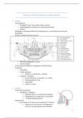
- RX OPG → welke structuren zien we?
1. fossa pterygopalatina
2. sinus maxillaris
3. mediale wand van de sinus
4. concha nasalis
5. fossae nasalia
6. neusseptum
7. neusbodem
8. arcus zygomaticus
9. capitulum articulare
10. tuber maxillae
11. processus styloideus
12. canalis N. Mandibularis
13. foramen mentale
14. linea obliqua externa
15. porus acusticus externus

o Effectieve stralingsdosis: 15 µSv
o Geen CI bij ZWS
- 3D reconstructie: effectieve stralingsdosis 100-500-700 µSv
- Anesthesie
o Zenuwblok
o Lokale anesthesie
▪ Xylocaïne – mepivacaïne – articaïne
o Anxiolyse – IV sedatie
▪ Cut-off meestal 2u → operatie < 2u: sedatie
▪ Monitoring BD & O2-sat
▪ Capnografie
▪ Patiënt is nuchter

2. Anatomie
- Cranio-faciaal evenwicht
o Mandibula = onderkaak
▪ Enige bot dat los is in cranio-faciale complex
▪ Ophanging met 2 condyli onder os temporale
▪ = onderste gelaatséénderde
- Mondopening
o Eerst rotatie (0 → 25mm), dan translatie (25 → 40mm)
▪ Rotatie tot je niet meer kan → kaakgewricht
naar voor → verder openen door translatie

1

, o Onderkaak scharniert op schedel
▪ = synoviaal gewricht met 2 soorten bewegingen
- Tandbogen: volwassenen
o 4 kwadranten
▪ 1 = Re boven 2 = Li boven
▪ 4 = Re onder 3 = Li onder
o 8 tanden/kwadrant
▪ Telling begint in het midden → achterste tand in kwadrant
• Bv. 1 = 1ste snijtand, 2 = 2de snijtand, 3 = hoektand
▪ Nummering
• 1ste cijfer = kwadrant
• 2de cijfer = tandnummer
• Bv. eerste molaar rechter bovenkaak = 16
▪ I = incisive = snijtanden
▪ C = canine = hoektanden
▪ P = premolaren
▪ M = molaren
o Distaal: alles naar posterieur vs.
mesiaal: alles naar interieur
o Vestibulair: richting vestibulum vs. linguaal: richting tong
o Labiaal: oppervlakte tegenover lip vs. bucaal: oppervlakte tegenover wang
- Tandbogen: kinderen
o 4 kwadranten
▪ 5 = Re boven 6 = Li boven
▪ 8 = Re onder 7 = Li onder

o 5 tanden/kwadrant
▪ Kinderen hebben geen premolaren
▪ I = 1ste melk snijtand
▪ II = 2de melksnijtand
▪ III = melkhoektand
▪ IV = 1ste melkmolaar
▪ V = 2de melkmolaar
- Gelaatspijlers
o Verticaal vs. horizontaal
o = stevig bot
o Tussen pijlers: luchthoudende ruimtes → kan heel dun bot zijn → breekt makkelijk


- Vascularisatie
o Frequente bloedingsplaatsen
▪ Neus: a. sphenopalatina
▪ Orbita

2

,- Innervatie
o n. V = n. trigeminus
▪ Sensibel: 2 takken
▪ Motorisch: 1 tak → bezenuwing kauwspieren
o n. VII = n. facialis
▪ Motorisch → bezenuwing mimische spieren
- Musculatuur
o 2 grote groepen parallel aan bezenuwing: mimische & kauwmusculatuur
▪ Aantal uitzonderingen
• Bv. m. digastricus: n V & VII, m. tensor tympani: n. V
o + tongbezenuwing
o Kauwspieren:
▪ Sluiten: m. temporalis, masseter, pterygoideus medialis
▪ Openen: m. pterygoideus lateralis, mylohyoideus, digastricus, geniohyoideus
▪ Sluiters sterker dan openers
▪ Cave: infectie → °trismus = spieren in spasme → °mond niet meer openen
- Anatomie van tand
o Zie Declerck
- Huidlijnen van Langer
o = plaats om in te snijden als toegang of excisie van
letsels op huid
o = zo esthetisch mogelijk

3. KO: intra-oraal
- Durf in mond kijken!
- Orale mucosa
o Soorten
▪ Vaste mucosa = gingiva = rond tanden & op verhemelte
• Vast, geen submucosa, lamina propria rechtstreeks op periost
▪ Losse mucosa: in wang & errond
• Beweeglijk, wel submucosa
▪ Gespecialiseerde mucosa: bv. op tong
o Kleur
▪ Graad van keratinisatie
• Hoe meer keratine, des te slechter onderliggende bloedvaten
zichtbaar zijn → blekere kleur
▪ Dikte van epitheel (bv. eelt onder prothese)
▪ Hoeveelheid melanine (bv. oraal melanoom)
▪ Concentratie & staat van dilatatie van capillairen in onderliggend
bindweefsel
o Opbouw
▪ Epitheel = epiderm van huid
▪ Lamina propria = dermis van huid
▪ Submucosa = subcutis van huid
▪ → geen duidelijke scheiding tussen lamina propria & submucosa


3

, o Letsels
▪ Letsel > 2w ter plaatse zonder genezingstendens → biopsie!
▪ Erosie = denudatie die boven basale laag van epitheel blijft → geen litteken
• Bv. pemphigus, erosieve lichen planus
▪ Ulcus = denudatie die doorheen basale laag van epitheel gaat
Erosie
• Bv. trauma, afte, virale infectie, mondkanker
▪ Papel = klein oppervlakkig verheven vast letsel < 1cm
diameter
Ulcus
• Bv. infectie, inflammatie, hyperplasie, neoplasie
Papel ▪ Plaque = vlak oppervlakkig verheven vast letsel van mucosa/huid > 1cm
diameter, kan dieper in dermis gaan
• Bv. witte letsels, roodwitte letsels, sommige
gepigmenteerde letsels
▪ Nodus = knobbeltje = vaste massa < 1cm diameter
• Gaat dieper in dermis dan papel Plaque
• Meestal los van overliggende mucosa/huid
▪ Tumor = diepe vaste massa
Nodus ▪ Cyste = holte, al dan niet met epitheel afgelijnd, kan vocht
bevatten
• Meestal in kaakbeen, ook in weke delen mogelijk Tumor


▪ Bulla = blaar, groter dan vesikel


a. TMJ (TEMPOROMANDIBULAIRE JOINT)
- Mogelijke symptomen
o Beweging
▪ Asymmetrie/bewegingsbeperking na trauma → beeldvorming
▪ Trismus = mondklem, kaakkramp = onmogelijkheid mond voldoende te
openen
• Door afwijking van kauwspieren of kaakgewricht
• Door mond- & gebitsontstekingen
o Eerder acuut opgetreden trismus
• → eigenlijk door alles wat prikkeling van spieren kan veroorzaken
o Geluid
▪ Knappende kaak = banaal → geruststelling
• Door dislocatie schijfje → mondopening: condyl verplaatst onder
schijfje → geluid
o Pijn
▪ Intra-articulair: bv. artrose
▪ Extra-articulair: bv. myogene overbelasting
▪ Gerefereerd: bv. uitstralende tandpijn

4

The benefits of buying summaries with Stuvia:

Guaranteed quality through customer reviews

Guaranteed quality through customer reviews

Stuvia customers have reviewed more than 700,000 summaries. This how you know that you are buying the best documents.

Quick and easy check-out

Quick and easy check-out

You can quickly pay through credit card or Stuvia-credit for the summaries. There is no membership needed.

Focus on what matters

Focus on what matters

Your fellow students write the study notes themselves, which is why the documents are always reliable and up-to-date. This ensures you quickly get to the core!

Frequently asked questions

What do I get when I buy this document?

You get a PDF, available immediately after your purchase. The purchased document is accessible anytime, anywhere and indefinitely through your profile.

Satisfaction guarantee: how does it work?

Our satisfaction guarantee ensures that you always find a study document that suits you well. You fill out a form, and our customer service team takes care of the rest.

Who am I buying these notes from?

Stuvia is a marketplace, so you are not buying this document from us, but from seller evdp. Stuvia facilitates payment to the seller.

Will I be stuck with a subscription?

No, you only buy these notes for $7.54. You're not tied to anything after your purchase.

Can Stuvia be trusted?

4.6 stars on Google & Trustpilot (+1000 reviews)

73918 documents were sold in the last 30 days

Founded in 2010, the go-to place to buy study notes for 14 years now

Start selling
$7.54  2x  sold
  • (0)
  Add to cart