This is a summary of the self-study particle for the course Neuropsychology and Psychopharmacology. It contains all the necessary information contained in the original document.
Neuropsy & Psychopharma
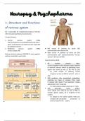
1. Structure and functions
of nervous system
NS = responsible for reception/processing of sensory
info from external/internal environments.
2 major divisions:
1. Central nervous system (CNS)
brain (completely surrounded/protected by skull) +
spinal cord (directly connected to brain/ protected
by vertebral column)
2. Peripheral nervous system (PNS) Red arrows → pathway by which CNS
nerves outside CNS communicates with sensory info
Black arrows → pathway by which the CNS
Arbitrary division between CNS/PNS → work together
communicates with somatic/automatic nervous
and are connected to each other.
system (2 divisions of PNS)
3 spec functions of NS:
1. NS receives sensory input.
sensory receptors in skin and other organs respond
to external/ internal stimuli by generating nerve
signals that travel by way of the PNS to CNS
vb: smell cookies → olfactory (smell)
receptors in nose use PNS to transmit info to
CNS
2. CNS performs info processing/ integration,
summing up input it receives from body
it reviews info, stores it as memories, creates
appropriate motor responses
- vb. Smell cookies evokes memories of taste
3. CNS generates motor output.
nerve signals from CNS go by way of PNS to
muscles, glands, organs, all in response of the
cookies.
- vb. Signals to the salivary glands make you
salivate/ CNS coordinates movement of
arms to reach for the cookie
1
, 3. Neuron anatomy
2. Nervous tissue
Sensory n takes nerve signals from a sensory
It contains 2 types of cells: receptor to the CNS.
1. Neurons - sensory receptors: special structures that
→cells that transmit nerve impulses between parts detect changes in environment.
of the NS Intern lies entirely within CNS. It receive input from
2. Neuroglia (or glial cells) sensory neurons and from other intern in CNS.
→ it support and nourish neurons. It outnr neurons They sum up all the info received from other
in brain. neurons before they communicate with motor n
several types of neuroglia in CNS with spec functions Motor n takes nerve impulses away from CNS to an
Microglia → phagocytic cells that help remove effector (muscle, fiber, organ, gland)
bacteria and debris - effectors carry out our responses to
Astrocytes → provide metabolic and structural environmental changes, whether these are ext-
support directly to the neurons internal
myelin sheath is formed from membranes of tightly Neurons vary in appearance, but all of them have 3
spiraled neuroglia distinct structures: a cell body, dendrites, axon
- Schwann cells (PNS) perform this function,
leaving gaps called nodes of ranvier Cell body contains nucleus and other organelles.
- Oligodendrocytes (CNS) form the myelin sheath Dendrites are short extensions that receive sign
from sensory receptors/ other neurons. Incoming
sign from dendrites can result in nerve sign that are
conducted by an axon.
Axon is the portion of neuron that conducts nerve
impulses. It can be long
- nerve fibers: individual axons and collectively
they form a nerve
In sensory n, a very long axon carries nerve sign from
dendrites associated with sensory receptor to CNS →
this exon is interrupted by cell body.
In intern/ motor n → multiple dendrites take signals to
the cell body, and then an axon conducts nerve sign
away from the cell body
= structure of sensory, inter-, motor neurons. The 3
types of neurons classified according to function
Sensory neuron → long axon covered by myelin
sheath that takes nerve impulses from dendrites to
CNS
Interneurons (in CNS) have short axon that isn’t
covered by myelin sheath
Motor neuro, → long axon covered by myelin
sheath that takes nerve impulses from CNS to an
effector
2
The benefits of buying summaries with Stuvia:
Guaranteed quality through customer reviews
Stuvia customers have reviewed more than 700,000 summaries. This how you know that you are buying the best documents.
Quick and easy check-out
You can quickly pay through credit card or Stuvia-credit for the summaries. There is no membership needed.
Focus on what matters
Your fellow students write the study notes themselves, which is why the documents are always reliable and up-to-date. This ensures you quickly get to the core!
Frequently asked questions
What do I get when I buy this document?
You get a PDF, available immediately after your purchase. The purchased document is accessible anytime, anywhere and indefinitely through your profile.
Satisfaction guarantee: how does it work?
Our satisfaction guarantee ensures that you always find a study document that suits you well. You fill out a form, and our customer service team takes care of the rest.
Who am I buying these notes from?
Stuvia is a marketplace, so you are not buying this document from us, but from seller mirtedeboeck. Stuvia facilitates payment to the seller.
Will I be stuck with a subscription?
No, you only buy these notes for $3.91. You're not tied to anything after your purchase.