Summary of Junqueira: Histology of the Nervous System with additional material from Netter's, DeFiore's and Weiss' Atlases. Includes abridged but detailed explanation of key points with Electron Microscopy, H&E, Immunohistochemistry and Diagrams.
02 connective tissue histology notes to 31 slides / images Class notes MTY1204 (histology) Junqueira's Basic Histology: Text and Atlas, Fiifteenth Edition, ISBN: 9781260026177
05 bone histology 39 notes to slides / images Class notes MTY1204 (histology) Junqueira's Basic Histology: Text and Atlas, Fiifteenth Edition, ISBN: 9781260026177
Immune System & Lymphoid Organs
All for this textbook (15)
Written for
David Tvildiani Medical University
Histology
All documents for this subject (7)
1
review
By: michelatornincasa • 4 year ago
Seller
Follow
galadashvili
Reviews received
Content preview
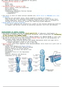
The Human Nervous System is composed of two parts:
• Central Nervous System or CNS
◦ Brain
◦ Brain Stem
◦ Spinal Cord
• Peripheral Nervous System or PNS
◦ Nerve Fibers- axons and dendrites
◦ Nerve Endings
◦ Peripheral Ganglia
◦ Subdivision: Autonomic Nervous System:
‣ Parasympathetic
‣ Sympathetic
• The cells in both of these nervous systems are: Nerve Cells or Neurons with long
processes
◦ Neurons are excitable cells, which respond to signals or Stimuli.
◦ They alter the ionic gradient/electrical potential that exists across their
plasma membranes, which leads to Membrane Depolarization and its propagation
through an Action Potential across the remaining plasma membrane.
• Glial Cells with short processes
◦ Glial Cells comprise the interstitial tissue of the nervous system and function
to support and protect neurons by providing neuronal nutrition and defense.
DEVELOPMENT OF NERVE TISSUE:
• The dorsal ectoderm, during the third week(14-16) of embryonic development is
subjected to signaling by the underlying notochord, which stimulates the thickening
of the ectoderm into NEURAL PLATE.
• The Neural plate forms a longitudinal Neural Groove with Neural Folds on each side.
• By day 24 the Neural Folds fuse forming a Neural Tube which is the primordial of
CNS: Cortex, Brain Stem and Spinal Cord.
◦ The Neural Tube contains Rostral and Caudal Neuropores which are open until:
‣ Rostral Neuropore closes on the 24-26 day
‣ Caudal Neuropore closes on the 27-28th day
• Neural Crest cells also separate from neuroectodermal cells which will give rise to
PNS components:
◦ Dorsal Root Ganglia of Spinal Nerves
◦ Ganglia of the Autonomic Nervous System
◦ Sensory Ganglia of Cranial Nerves
◦ Meninges of the Brain and Spinal Cord
◦ Melanocytes
◦ Chromaffin Cells of the Suprarenal
Medulla
◦ Somites: Skeletal and Muscular
components of the Head
, Mes
Met alphabetical
Myl
NEURONS:
• Neurons are signal transmitting functional units of both CNS and PNS:
• They are composed of three main parts:
◦ Cell Body/Soma:
‣ Unusually large and Spherical.
‣ Contains Euchromatic Nucleus and
Prominent Nucleolus(one or more)
• The nucleus is large compared to the
cytoplasm/perikaryon
‣ The cytoplasm contains Basophilic
Chromatophilic Substance of Nissl Bodies
which are clumps of highly developed
flattened cisternae of RER containing
Polyribosomes in between them which
synthesize proteins for internal use and
export.
• Nissl Substance is only found in the
perikaryon
‣ Highly developed Golgi Complex with
multiple stacks of flattened sacs and
associated vesicles and vacuoles can also be
seen which is responsible for packaging and
concentration of secretary products, post
translational modification of macromolecules
and supply of lysosomes which accumulate
lipofuscin pigment in long-lived neurons. as
well as Golgi apparatus.
◦ Dendrites:
‣ Numerous elongated processes extending from the
Perikaryon/Cytoplasm.
‣ They consist of a thick base, which
branches out and ends in thin processes
called Dendritic Spines(gemmules).
Dendritic spines are where synapses from
other neurons make contact with the cell
thus making dendrites the principal site
for signal reception and processing.
‣ Dendritic Spine are associated with Neural
Plasticity which underlies our ability to
adapt, learn and have memory.
‣ Neurons are covered with many dendrites
increasing the number of axons it can receive.
• Purkinje Cells of cerebellum receive up to 200,000 functional axon synapses.
◦ Axons:
‣ Axons are fine long(100cm) cylindrical processes with a constant diameter.
‣ They originate from Axonal Hillocks and are composed of:
• Axolemma which is its plasma membrane
• Axoplasm which contains mitochondria, microtubules, microfilaments, and
SER.
◦ Microtubules are of special importance as they provide intracellular
The benefits of buying summaries with Stuvia:
Guaranteed quality through customer reviews
Stuvia customers have reviewed more than 700,000 summaries. This how you know that you are buying the best documents.
Quick and easy check-out
You can quickly pay through credit card or Stuvia-credit for the summaries. There is no membership needed.
Focus on what matters
Your fellow students write the study notes themselves, which is why the documents are always reliable and up-to-date. This ensures you quickly get to the core!
Frequently asked questions
What do I get when I buy this document?
You get a PDF, available immediately after your purchase. The purchased document is accessible anytime, anywhere and indefinitely through your profile.
Satisfaction guarantee: how does it work?
Our satisfaction guarantee ensures that you always find a study document that suits you well. You fill out a form, and our customer service team takes care of the rest.
Who am I buying these notes from?
Stuvia is a marketplace, so you are not buying this document from us, but from seller galadashvili. Stuvia facilitates payment to the seller.
Will I be stuck with a subscription?
No, you only buy these notes for $2.99. You're not tied to anything after your purchase.