100% satisfaction guarantee Immediately available after payment Both online and in PDF No strings attached
logo-home
Summary Histology of Muscle Tissue $2.99   Add to cart

Summary

Summary Histology of Muscle Tissue

 165 views  4 purchases
  • Course
  • Institution
  • Book

Summary of Junqueira: Histology of the Muscle with additional material from Netter's, DeFiore's and Weiss' Atlases. Includes abridged but detailed explanation of key point with Electron Microscopy, H&E, Immunohistochemistry and Diagrams.

Preview 2 out of 9  pages

  • No
  • Chapter 10
  • February 2, 2020
  • 9
  • 2019/2020
  • Summary
avatar-seller
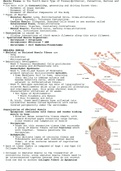
Muscle Tissue is the fourth Basic Type of Tissue(Epithelial, Connective, Nervous and
Muscle)
• Its main role is Contractility, generating and driving forces that:
◦ Movement of Organ Systems
◦ Movement of Blood
◦ Movement of Skeletal Components of the Body
• Types of Muscle:
◦ Skeletal Muscle: Long, Multinucleated Cells, Cross-striations,
‣ Quick, Forceful, Voluntary Contractions.
◦ Cardiac Muscle: Elongated, Branched Cells bound to one another called
Intercalated Discs, Cross-striations
‣ Rhythmic, Vigorous, Involuntary Contractions
◦ Smooth Muscle: Fusiform Cells, Lacking cross-striations
‣ Slow, Involuntary Contractions.
• Contraction is caused by:
◦ Sliding interactions of thick myosin filaments along thin actin filament.
• Specialized Muscle Organelles:
◦ Sarcoplasm = Cytoplasms
◦ Sarcoplasmic Reticulum = SER
◦ Sarcolemma = Cell Membrane/Plasmalemma

SKELETAL MUSCLE
• Skeletal or Striated Muscle Fibers are
◦ Long
◦ Cylindrical
◦ Multinucleated
• Embryologic Development:
◦ Week 4 Paraxial Mesodermal Cells proliferate
and elongate and differentiate into
Mesenchymal Myoblasts.
◦ Week 5, end-to-end fusion of Myoblasts
produce syncytial multinucleated myotubes.
‣ Some Myoblasts fail to fuse, retaining
their ability to undergo mitosis and
become Satellite cells, which remain
adjacent to differentiated fibers.
◦ Week 9, Myotubes synthesize two sets of longitudinally
oriented Myofilaments which align in parallel alternating
and overlapping rays, this gives skeletal muscle
characteristic striations at 20 weeks.
‣ Two Types of Myofilaments:
• Thick Filaments are Myosin
• Thin Filaments are Actin
◦ At birth, these myofilaments have collected into
cylindrical bundles called Myofibrils with nuclei
located peripherally under the sarcolemma.

Organization of Skeletal Muscle
◦ Epimysium: continuous with fascia and tendon binding
muscle to bone
‣ External dense connective tissue sheath, with
inward directed septa containing large nerves,
blood vessels and lymphatics of muscle
◦ Perimysium:
‣ Dense but thin connective tissue that surrounds
a group of muscle fibers called Fascicles which
work together. Vessels penetrate the
Perimysium.
◦ Endomysium:
‣ Thin delicate layer of reticular fibers and
fibroblasts surround each individual muscle
fiber, it also contains network of capillaries
that supply O2.
• Each of these layers is composed of Type I and Type III
Collagen(Reticulin)

• Collagen in Skeletal Muscle is the transmitter of
contractile force, since skeletal muscle fibers need to
be interconnected together because they do not stretch
from one end to another Collagen Fibers in Epimysium

, connect with Collagen fibers
of the Tendon of the next
muscle.

Organization of Skeletal
Muscles Fibers
• Muscle Fiber is composed of
several parallel Myofibrils
◦ Myofibrils consist of end
to end registrations of
Sarcomere, functional
subunit of contractile
apparatus, which give it
distinctive pattern of
transverse striation.
• Each Sarcomere Unit consists of Myofilaments:
◦ A Bands: Anisotropic Bands that are
birefringent in polarized light microscopy
giving a DARK BAND
‣ Within each A Band, there is an less
electron dense H zone that appears paler,
within the H zone there is an M Line(Mitte,
middle) which contains a myosin binding
protein which holds thick filaments in
place called Myomesin and Creatine Kinase
an enzyme which mediates the transfer of
Phosphate group from Creatine Phosphate to
ADP to provide ATP when energy demand in
muscle is high.
◦ I Bands: Isotropic Bands that do not alter
polarized light giving LIGHT BANDS
‣ Each I Band is transversed by a Z
Disc(zwischen, between)
• A Sarcomere Unit Extends from Z Disc to
Z Disc.
◦ The I and A banding is due to Myofilaments
‣ Myofilaments are attached to the Zwischen Disc by two proteins
• a-actinin and nebulin anchor Thin Actin myofilaments
• Titin the largest protein in the body(3700kd) anchoring Thick Myosin
myofilaments
‣ Thick Filaments: Contain Molecules of Myosin and produce Dark A bands
• Myosin Contains 2 Heavy Chains that are Twisted Together as a Myosin Tail
and act as Motor Proteins, and 2-4 Light Chains form the Head of the
Myosin Molecule that Binds ATP and Myosin Binding Site on Thin Filaments
forming crossbridges.
‣ Thin Filaments: Contain Molecules of F-actin(composed of G Actin), Troponin,
Tropomyosin.
• Thin Filaments run in
between thick filament.
• Each G-actin component of
F-actin has a binding site
for Myosin Heads.
• Tropomyosin is a coil of
two polypeptides wrapping
around in a groove between
two twisted Actin Chains.
• Troponin composed of three
subunits:
◦ TnT binding
Tropomyosin
◦ TnC binding Calcium
◦ TnI regulating
actin-myosin
interactions.

The benefits of buying summaries with Stuvia:

Guaranteed quality through customer reviews

Guaranteed quality through customer reviews

Stuvia customers have reviewed more than 700,000 summaries. This how you know that you are buying the best documents.

Quick and easy check-out

Quick and easy check-out

You can quickly pay through credit card or Stuvia-credit for the summaries. There is no membership needed.

Focus on what matters

Focus on what matters

Your fellow students write the study notes themselves, which is why the documents are always reliable and up-to-date. This ensures you quickly get to the core!

Frequently asked questions

What do I get when I buy this document?

You get a PDF, available immediately after your purchase. The purchased document is accessible anytime, anywhere and indefinitely through your profile.

Satisfaction guarantee: how does it work?

Our satisfaction guarantee ensures that you always find a study document that suits you well. You fill out a form, and our customer service team takes care of the rest.

Who am I buying these notes from?

Stuvia is a marketplace, so you are not buying this document from us, but from seller galadashvili. Stuvia facilitates payment to the seller.

Will I be stuck with a subscription?

No, you only buy these notes for $2.99. You're not tied to anything after your purchase.

Can Stuvia be trusted?

4.6 stars on Google & Trustpilot (+1000 reviews)

64438 documents were sold in the last 30 days

Founded in 2010, the go-to place to buy study notes for 14 years now

Start selling
$2.99  4x  sold
  • (0)
  Add to cart