100% satisfaction guarantee Immediately available after payment Both online and in PDF No strings attached
logo-home
Casusbundel (casus 1-13) $6.44   Add to cart

Case

Casusbundel (casus 1-13)

 2 views  0 purchase
  • Course
  • Institution

Alle casussen in het blok Circulatie en Ademhaling I

Preview 4 out of 83  pages

  • November 11, 2024
  • 83
  • 2022/2023
  • Case
  • K. hendriks
  • 7-8
avatar-seller
CIRCULATIE & ADEMHALING
JAAR 1 – CASUS 1 T/M 13


Femke van Heel

,Casus 1 Inspanning

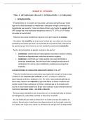
Wat is de anatomie van het hart? (macroscopisch, ook kransslagadersysteem)

, Het hart bevindt zich in het mediastinum (middelste holte van de thorax), en loopt
schuin uit van de 2e rib tot de 5e intercostaalruimte
 Pericardium: dubbelwandige zak dat het hart omvat
o Pericardium fibrosum: bovenste laag van het pericardium, zit los om het hart
heen. Het bestaat uit een stevig bindweefsel en dient hierdoor voor
bescherming van het hart. Het houdt het hart op zijn plek en voorkomt dat het
hart te vol loopt met bloed. Het pericardium fibrosum zit deels vast aan het
diafragma
o Pericardium serosum: onderste laag van het pericardium, vormt een
gesloten zak om het hart. Het bestaat uit een dun, glad, tweelaags membraan.
Het pericardium serosum bestaat dus uit drie delen:
 Pariëtale pericardium: buitenste laag, ondoordringbaar en erg stevig.
De laag scheidt aan de binnenkant in de pericardiale ruimte een
vloeistof af, waardoor de twee lagen van het pericardium makkelijker
over elkaar heen glijden tijdens het pompen van het hart. De
onderkant van deze laag is samengesmolten met de centrale pees
van het diafragma. Hierdoor heeft de ademhaling invloed op de vulling
van het hart

,  Pericardiale ruimte: ruimte tussen de pariëtale en viscerale laag die
met vloeistof gevuld is (door het pariëtale pericardium)
 Viscerale pericardium: binnenste laag van het pericardium serosum,
deze is erg dun en vergroeid met het hart
o Het pericardium ligt achter het sternum (borstbeen), van de 2e tot de 6e rib en
de Th5-Th8 wervels
 Apex/hartpunt: inferieure kant van het hart, steekt schuin naar links voor
 Basis: superieure kant van het hart, waar de grote arteriën en venen zitten. Het wordt
gevormd door het linker en rechter atrium
 Grote belangrijke vaten:
o Truncus pulmonaris: met aftakkingen a. pulmonalis dexter en sinistra
o Arcus aortae: met aftakkingen truncus brachiocephalicus, a. carotis
cummonis sinistra en a. subclavia sinistra
o Vena cava superior
o Vena cava inferior
 Hartkleppen:
o Valva atrioventricularis sinistra (mitralisklep): tussen het linker atrium en
het linker ventrikel. Dit is de enige klep met twee klepbladen, de andere
kleppen hebben drie klepbladen
o Valva atrioventricularis dextra (tricuspidalisklep): tussen het rechter
atrium en het rechter ventrikel. Zorgt ervoor dat er geen bloed terugstroomt
van het rechter ventrikel naar het rechter atrium
o Valva aortae (aortaklep): tussen het linker ventrikel en de aorta
o Valva trunci pulmonalis (pulmonalisklep): tussen het rechter ventrikel en
de truncus pulmonaris (longslagader)
 De aortaklep en de pulmonalisklep zijn halvemaanvormige kleppen
(valvae semilunares)


 Rechter atrium: bestaat uit anterieure en posterieure kant, die worden gescheiden
door een richel genaamd het crista terminalis
o Ontvangt bloed vanuit de vena cava inferior en de sinus coronarius. Dit is
een grote ader, onderdeel van het kransslagadersysteem, die al het

The benefits of buying summaries with Stuvia:

Guaranteed quality through customer reviews

Guaranteed quality through customer reviews

Stuvia customers have reviewed more than 700,000 summaries. This how you know that you are buying the best documents.

Quick and easy check-out

Quick and easy check-out

You can quickly pay through credit card or Stuvia-credit for the summaries. There is no membership needed.

Focus on what matters

Focus on what matters

Your fellow students write the study notes themselves, which is why the documents are always reliable and up-to-date. This ensures you quickly get to the core!

Frequently asked questions

What do I get when I buy this document?

You get a PDF, available immediately after your purchase. The purchased document is accessible anytime, anywhere and indefinitely through your profile.

Satisfaction guarantee: how does it work?

Our satisfaction guarantee ensures that you always find a study document that suits you well. You fill out a form, and our customer service team takes care of the rest.

Who am I buying these notes from?

Stuvia is a marketplace, so you are not buying this document from us, but from seller femkevanheel. Stuvia facilitates payment to the seller.

Will I be stuck with a subscription?

No, you only buy these notes for $6.44. You're not tied to anything after your purchase.

Can Stuvia be trusted?

4.6 stars on Google & Trustpilot (+1000 reviews)

67474 documents were sold in the last 30 days

Founded in 2010, the go-to place to buy study notes for 14 years now

Start selling
$6.44
  • (0)
  Add to cart