Cardiovascular Disease Summary
ENDOTHELIAL CELL FUNCTION → all CVDs contain some form of endothelial dysfunction
Cellular plasticity:
- heterogeneity of the vasculature: the
capillary network is different between organs
→ the blood supply is depended on the
metabolism of the tissue/organ
- organ-specific EC phenotypes
- endothelial-mesenchymal transition
Functional plasticity:
- barrier function (adhesion molecules):
1. Tight junctions (occludins): sealing of
the EC layer (membrane fusion)
2. Adherence junctions (VE-cadherin):
cell-cell connections (distribution of force), gap junctions (EC-EC communication (connection of
cytoplasm))
→ formation of gradients (pressure is high in vasculature, low in tissue, metabolites (active transport
from blood into tissues), inhibition of leukocyte migration
- immunology (leukocyte migration)
- thrombogenicity: heparan-sulfaphates (inhibition of thrombosis), secretion of coagulation factors
(PAI1/tPa/vWF), inhibition of platelet aggregation (NO/PGI2)
- vasomotor function (NO, ET1)
- vascularisation (vasculogenesis, angiogenesis)
VASCULAR SYSTEM
Function: transport of O2, nutrients, hormones and waste products
Vessel types: aorta → arteries → arterioles → capillaries → venules → veins → vena cava
Structure of vessels:
Tunica Externa Lamina Tunica Media Lamina Tunica Intima
(/Adventitia) elastica elastica
Cell type Fibroblasts externa Smooth muscle interna Endothelial cells
Matrix Collagens (ECM) Elastin fibers Basal membrane
(collagen I) (collagen III +
elastin)
Function Stiffness, Vasoconstriction Coagulation,
anchoring to or vasodilation transport,
tissue inflammation
Distribution of blood over smaller blood vessels → increases the cross-sectional area, flow rate ↓,
diffusion (gas exchange rate) ↑
Development of the vascular system (takes 6 weeks)
- day 12 post-fertilization: start
- first development of vasculature, then development of heart
- blood islands → extraembryonic vasculature (placenta, umbilical cord)
- mesoderm → embryonic vasculature
- 8 weeks post-fertilization: complete functional CV system
,Mesoderm formation patterning: induced by cell-cell interaction
Blood vessel formation: BMP4 → mesoderm differentiation → FGF-2 → hemangioblast formation
→ (proliferation, FGF-2) → blood island
→ (differentiation) → angioblasts (blood vessel-forming cells), hematopoietic stem cells, mural cells
(pericytes/SMCs)
Vasculogenesis: formation of a primary vascular plexus in the embryo proper and extra-embryonic
tissues
- induced by VEGF/VEGFR signalling
- VEGF-A induces the formation of a capillary plexus
- mesoderm → hemangioblasts → tube formation → primary capillary plexus
- regulation is largely unknown
- mutations in VEGF/VEGFR/co-receptors → embryonic lethality ↑
Genes*
VEGF-A Vasculogenesis, angiogenesis
VEGF-B Embryonal angiogenesis
VEGF-C Lymph angiogenesis
VEGF-D
VEGF-E Viral-derived VEGF, function unknown
Receptors
VEGFR1 Angiogenesis (stalk cell differentiation)
VEGFR2 Vasculogenesis, angiogenesis (tip cell differentiation)
Co-receptors: NRP1 & NRP2: amplify VEGFR2 signalling
VEGFR3 Lymph angiogenesis
*VEGF-A(-E) isoforms: differences in heparin binding domain (binding to ECM) → presence
determines the diffusion capacity of VEGF (long versus short distance)
Arteriovenous differentiation
- starts before blood flow starts: important genetic component,
influenced/strengthened by circulation (mechanical forces)
- aorta & vena cava originate from the same primitive vessel
- ephrins (= ligand) → arteries
- ephs (= receptor) → veins
- bidirectional signalling
- cell-cell contact required, but results in repulsion
1. Arterial identity (active process)
- VEGF/VEGFR2 → EphB4 ↑
- Notch1 → EphrinB2↑, EphB4↓ (EphB4 expression is inhibited)
2. Venous identity (passive process)
- VEGF/VEGFR2 → EphB4 ↑
- segregation: active process (EphB4 signalling results in migration (away from aorta))
, Tip cell Stalk cell Phalanx EC
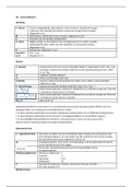
Location At the tip of the sprout Behind tip cell In blood vessel
Polarisation High; filopodia None None
Receptor expression VEGFR2-3, NRP1, DLL4 VEGFR1-2, Notch1/4 VEGFR1-2, Tie2
MMP expression Yes, MT1-MMP (col. IV) No No
Lumen None Developing Yes
Proliferation, rate No Yes, high Yes, low
Angiogenesis: formation of new blood vessels from existing ones
- some cells divide, some cells migrate
1. Tip cell
- VEGF binds to VEGFR2/3 → tip cell differentiation → expression of DII4 → DII4/Notch1 interaction
inhibits tip cell differentiation of adjacent cells by the inhibition of VEGFR2 expression
- filopodia/lamellopodia: high expression of VEGFR2 and integrins (sense gradients)
- VEGF/VEGFR2 → FAK activation → actin polymerization → movement
- MT1-MMP (MMP14): matrix metalloprotease that degrades the basal lamina (ECM) → allows
migration
2. Stalk cell
- DII4/Notch1 interaction inhibits VEGFR2/3 expression in stalk cells → decreased sensitivity to VEGF
- Notch1 → activates VEGFR1 (Flt1) expression → proliferation ↑
- Notch1 → activates integrin expression → cell-matrix interaction = stabilization
- Notch1 → activates Wnt signalling → cell-cell contacts ↑ (stabilization)
- pinocytosis (formation and endocytosis) of membrane intrusions → vacuole formation →
coalescence of vacuoles → intracellular lumen → fusion of intercellular lumens (ECs share 1 lumen,
but their cytoplasm remains separated)
3. Phalanx EC
- non-proliferating endothelium
- strong cell-cell adhesions of VE-Cadherin homodimers and Tie2-Ang1-Tie2 binding
- low VEGFR1/VEGFR2 expression (barely enough for cell survival)
- sVEGFR1: secreted decoy receptor for VEGF-A
Mural cells: form the tunica media and surround the endothelium
- SMCs: outside the basement membrane, multicellular layer
- pericytes: inside the basement membrane, single cells or single cell layer
- PDGF-BB: dimer that recruits mural cells (PDGFRβ), produced by tip cell, binds to the ECM
- Ang1 of mural cell activates Tie2 on EC → 1. PI3K-Akt → cell survival
2. NFκB inhibition → no inflammation
3. Rho kinase inhibition → EC-EC interactions are
stabilized with adherence junction (VE-Cadherin)
- S1P (mural cell) activates S1PR1 (EC) → activates Rac signalling → promotes EC-EC interactions (VE-
Cadherin) and EC-pericyte interactions (N-Cadherin)
- TGFβ (EC) activates TGFBR1(ALK5) (mural cell) → activates SMAD2/3 signalling → ECM production
(stabilization), SMC differentiation (contraction), inhibits mural cell proliferation (stabilization)
Lymph angiogenesis
- lymph vessels differentiate from the venous endothelium
- starts with blood clot (has no anti-thrombosis capacity)
, - lymph angiogenesis follows a program similar to sprouting angiogenesis, BUT
- VEGF-C (not VEGF-A) induces lymph angiogenesis
- VEGF-C signals via VEGFR3 (not VEGFR1)
- valves
ANGIOADAPTATION
- guarantee the supply of nutrients and oxygen, quick responses
- blood pressure → regulation of diffusion volume
- blood flow → regulation of diffusion time
- vascularization → regulation of diffusion capacity
- changes are made constant and are on demand
- remodelling to deal with metabolic demand, regulate blood flow to different organs, remove non-
function and excessive blood vessels
Arterial blood pressure is regulated autonomously (independent of blood flow and cardiac output)
- short term regulation: changes in volume distribution / cardiac output
- long term regulation: renin-angiotensin-aldosteron system (RAAS)
HIF1α signalling
- hypoxia induced → vasodilatation,
angiogenesis, oxygen transport capacity
→ increased blood flow (speed), flow
volume, increased transport capacity
Mechanotransduction: conversion of
mechanical signals to biochemical
signals → gene expression
- shear stress: increases during
increased blood flow
- normal stress: increases during
increased blood pressure
- no/limited blood flow = loss of shear stress → angiopoietine-2 activation → inhibits Tie2
Collateral formation: redistribution of flood flow by the arterialisation of a capillary or ateriole
- induced by a (massive) increase in shear stress
- can originate from newly-formed blood vessels (ischemia driven) or from pre-existing blood vessels
(flow driven)


