100% satisfaction guarantee Immediately available after payment Both online and in PDF No strings attached
logo-home
Samenvatting Echo leerdoelen uitgewerkt P5 $5.37
Add to cart

Summary

Samenvatting Echo leerdoelen uitgewerkt P5

 60 views  3 purchases
  • Course
  • Institution
  • Book

in dit document zijn de leerdoelen van echografie periode 5 uitgewerkt

Preview 2 out of 7  pages

  • Unknown
  • August 20, 2020
  • 7
  • 2019/2020
  • Summary
avatar-seller
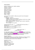
vEC leerdoelen periode 5

Anatomie van de lever
 De ligging en (topografische) anatomie van de lever beschrijven:
o Linker leverkwab (lobus sinister)
o Rechter leverkwab (lobus dexter)
o Lobus caudatus → ligt craniaal
o Lobus quadratus
o Ligamentum teres (afgrenzing van lobus quadratus)
o Interlobaire fissuur (afgrenzing van lobus caudatus)
o Ligamentum falciforme (niet te zien op de echo → in anatomie de scheidingslijn
tussen dextra en sinistra)
o Leverpoort/hilus;


 Op het echografische beeld van de lever de structuren benoemen en aanwijzen;
1. Vena portae ramus sinistra
2. Hart
3. Vena hepatica media
4. Vena cava inferior Sagittaal
5. Confluens Linker leverkwab
6. Arteria hepatica communis
7. Lobus caudatus
8. Interlobaire fissuur
1. Rectus abdominalis/buikspier
2. Ligamentum teres
Transversaal
3. Venae porta ramus sinistra
4. Vena cava inferior
5. Vena hepatica media
6. (Interlobaire fissuur)/slagschaduw van ligamentum teres
7. Pancreas cauda
8. Aorta abdominalis

, 1. Caudale leverpunt
2. Diafragma
3. Rechter nier
4. Rechter leverkwab Sagittaal


1. Vena porta takje
2. Vena hepatica dextra
3. Vena hepatica dextra
4. Rechter leverkwab Transversaal
5. Vena hepatica media
6. Vena cava inferior
7. Diafragma

1. galblaas
2. vena portae intrahepatisch
3. rechter leverkwab
4. ligamentum teres Oblique
5. lobus quadratus
6. vena cava inferior

1. Latero-ventrale leverrand
Oblique
2. Diafragma (lateraal)
Vena hepatica dextra
3. Vena portae ramus dextra
4. Spiegelartefact (niet duidelijk,
wel plaats waar vaak zichtbaar)/lon


 Aan de hand van een echografische opname beschrijven in welke richting de genummerde
structuren/ vaten zijn afgebeeld.

 Verschillende systemen in de lever benoemen
o Portale (bloedvaten) systeem
 Vena porta voert bloed af met
afvalstoffen de lever in
o Veneuze systeem
 Loopt af naar craniaal, voeren
af naar de vci
o Arteriële systeem
 Voert zuurstofrijk bloed aan
o Galwegen
 Stroom de lever uit, gal moet
naar de darmen

The benefits of buying summaries with Stuvia:

Guaranteed quality through customer reviews

Guaranteed quality through customer reviews

Stuvia customers have reviewed more than 700,000 summaries. This how you know that you are buying the best documents.

Quick and easy check-out

Quick and easy check-out

You can quickly pay through credit card or Stuvia-credit for the summaries. There is no membership needed.

Focus on what matters

Focus on what matters

Your fellow students write the study notes themselves, which is why the documents are always reliable and up-to-date. This ensures you quickly get to the core!

Frequently asked questions

What do I get when I buy this document?

You get a PDF, available immediately after your purchase. The purchased document is accessible anytime, anywhere and indefinitely through your profile.

Satisfaction guarantee: how does it work?

Our satisfaction guarantee ensures that you always find a study document that suits you well. You fill out a form, and our customer service team takes care of the rest.

Who am I buying these notes from?

Stuvia is a marketplace, so you are not buying this document from us, but from seller kimmbrt. Stuvia facilitates payment to the seller.

Will I be stuck with a subscription?

No, you only buy these notes for $5.37. You're not tied to anything after your purchase.

Can Stuvia be trusted?

4.6 stars on Google & Trustpilot (+1000 reviews)

52510 documents were sold in the last 30 days

Founded in 2010, the go-to place to buy study notes for 14 years now

Start selling
$5.37  3x  sold
  • (0)
Add to cart
Added