scope and definition of biophysics and its general application
basic biophysic mechanisms
Connected book
Book Title:
Author(s):
Edition:
ISBN:
Edition:
More summaries for
Biophysics, Foundation of Biophysics (32)
All for this textbook (2)
Written for
EGERTON UNIVERSITY
Introduction to Biophysics (317)
All documents for this subject (1)
Seller
Follow
GRADUATEnurse
Reviews received
Content preview
EU 2021 BMED 317
BIOPHYSICS
Definition
Biophysics is a bridge between biology and physics.
Biophysics is an interdisciplinary science which uses methods and theory of physics to study biological
system. It spans from the molecular scale to whole organisms and ecosystems. Biology studies life in its
variety and complexity. It describes how organisms go about getting food, communicating, sensing the
environment and reproducing. On the other hand, physics looks for mathematical laws of nature and
makes detailed prediction about the forces that drive idealized system. Biophysics fuses the complexity of
life and simplicity of physical laws. Therefore, biophysics is a bridge of biology and physics.
Biology studies life in its variety and complexity. It describes how organisms go about getting food,
communicating, sensing the environment, and reproducing. On the other hand, physics looks for
mathematical laws of nature and makes detailed predictions about the forces that drive idealized systems.
Spanning the distance between the complexity of life and the simplicity of physical laws is the challenge
of biophysics. Looking for the patterns in life and analyzing them with math and physics is a powerful
way to gain insights.
Biophysics looks for principles that describe patterns. If the principles are powerful, they make detailed
predictions that can be tested.
I. Role of Biophysics in revealing the structure of DNA
DNA, unlike proteins, is an exceedingly large molecule which does not lend itself to crystallisation. It is
usually extracted in the form of the sodium salt (to neutralise the negatively charged phosphates) to yield
a highly viscous suspension of molecular fibers. These fibers are disorderly compared with single crystals
and produce smears rather than spots. It was in Maurice Wilkins lab that
techniques were developed to form thin fibers of DNA. These almost
invisible, spiderweb-like filaments in which the DNA molecules were
deduced to be neatly aligned alongside one another. This technique formed
the basis for studying the X-ray patterns formed by DNA fibers kept at high
humidity. Such fibers produce diffraction patterns that hint at the clarity
obtainable from a perfect single crystal. This patterns is visualised of many
pictures of a crystal taken at different angles, but with sorted out the
overlapping aspects. This was a very hard proces. Rosalind Franklin joined
Wilkins lab and further developed the technique and procedures to produce
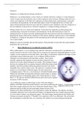
diffraction pictures of outstanding quality. One of those pictures is called
Photo 51. The data she obtained enabled Watson and Crick to piece together
the puzzle of DNA structure.
X-ray diffraction studies on DNA began in June 1950 when Maurice
Wilkins asked PhD student Raymond Gosling to assist him in diffracting the
DNA fibre samples prepared by the Swiss biochemist, Rudolf Signer. Fibre
diffraction did not usually provide good quality images because of the
thinness of the fibres and therefore a very small mass to scatter the radiation. Nevertheless, the fibres’
remarkable uniformity when wetted allowed Wilkins to manipulate them into a bundle and mount them
on a wire frame to obtain x-ray diffraction images. The initial images showed promise but Wilkins and
Gosling were greatly assisted by J T Randall’s own experience with X-ray diffraction. He advised how
the surrounding air could affect the x-ray scattering. The solution was to pass hydrogen through the
1
, EU 2021 BMED 317
camera and control the relative humidity of the sample. With this in place, the resulting images were
much sharper and showed a clear crystalline diffraction pattern.
It was in late 1950 that the theoretical physicist Alec Stokes first noticed an interesting observation from
the images. He realised that there was no diffraction at all along the length of the molecules: a sign that
DNA might be helical. However, the King’s College team needed far sharper images to confirm this
hypothesis. This required a new X-ray camera that could work on single fibres. Through a fortunate
coincidence, Werner Ehrenberg and W E Spears had just developed one at Birkbeck: this was generously
loaned to the King’s College team.
Before the new camera was set up, it was decided that Rosalind Franklin, who was joining the laboratory
from Paris, would replace Wilkins in producing the x-ray diffraction images with the continued assistance
of Raymond Gosling. Both Stokes and Wilkins continued working on the problem with the latter
embarking on some rough tests with the old X-ray diffraction camera on various DNA specimens that
produced an observed “X” crossed pattern. The X pattern of diffraction was created by the x-ray radiation
scattering at right angles off the "zigzag" structure of the DNA chain. This interpretation was further
supported when Franklin and Gosling produced the first “B” structure X-ray patterns in the late summer
of 1951. This was a crucial development as it showed two observed states of DNA: crystalline “A” and
semi-crystalline “B” (the best B structure diffraction photograph became known as “Photo 51”).
X-ray diffraction (or X-ray crystallography) was the chief physical method used to determine the structure
of DNA.
X-ray diffraction is the method of projecting a beam of X-ray radiation at a target object and through to a
photographic film on the far side. A series of spots appear on the photographic film following this
exposure, which is formed by the x-ray radiation diffracting off the structure that they passed through.
These diffraction patterns give an indication of the general structure of the object (such as an inorganic
crystal or macro- molecule such as DNA) which can then be delineated using complex mathematical
formulas.
The reason why X-ray beam is required in the first place is that atoms are too small (0.1nm between them,
bearing in mind that 1 millimetre = 1000000 nanometres) to be revealed using visible light and therefore
could not be viewed by a light microscope (even an electron microscope does not possess the required
magnification). X-ray radiation fits the appropriate wavelength to be diffracted by the object and produce
visible results.
What the X-ray beam are diffracting is not the entire atom but the orbiting electrons that are close enough
to the nucleus of the atom to give a good indication of the structure of the unit cel. The end image is
known as an electron density map of that unit cell. However due to the incredibly weak image a single
molecule would produce, a crystalline structure is used instead, for example common salt (NaCl), since a
crystalline structure provides a huge number of molecules arranged in the same orientation and therefore
produces the same scattering effect on the X-ray beams.
Bragg’s Law
The law states that when the x-ray is incident onto a crystal surface, its angle of incidence, θ, will reflect
back with a same angle of scattering, θ. And, when the path difference, d is equal to a whole
number, n, of wavelength, a constructive interference will occur.
The general relationship between the wavelength of the incident X-rays, angle of incidence and spacing
between the crystal lattice planes of atoms is known as Bragg's Law, expressed as:
2
The benefits of buying summaries with Stuvia:
Guaranteed quality through customer reviews
Stuvia customers have reviewed more than 700,000 summaries. This how you know that you are buying the best documents.
Quick and easy check-out
You can quickly pay through credit card or Stuvia-credit for the summaries. There is no membership needed.
Focus on what matters
Your fellow students write the study notes themselves, which is why the documents are always reliable and up-to-date. This ensures you quickly get to the core!
Frequently asked questions
What do I get when I buy this document?
You get a PDF, available immediately after your purchase. The purchased document is accessible anytime, anywhere and indefinitely through your profile.
Satisfaction guarantee: how does it work?
Our satisfaction guarantee ensures that you always find a study document that suits you well. You fill out a form, and our customer service team takes care of the rest.
Who am I buying these notes from?
Stuvia is a marketplace, so you are not buying this document from us, but from seller GRADUATEnurse. Stuvia facilitates payment to the seller.
Will I be stuck with a subscription?
No, you only buy these notes for $9.99. You're not tied to anything after your purchase.