100% satisfaction guarantee Immediately available after payment Both online and in PDF No strings attached
logo-home
Summary 2.4. Problem 2: Connections $3.44   Add to cart

Summary

Summary 2.4. Problem 2: Connections

2 reviews
 58 views  4 purchases
  • Course
  • Institution
  • Book

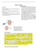
This is a detailed summary of problem 2 for course 2.4 Perception.

Preview 2 out of 17  pages

  • No
  • 3rd edition, chapter 4, pages 142-145, pages 177-206
  • September 1, 2021
  • 17
  • 2020/2021
  • Summary

2  reviews

review-writer-avatar

By: aronkoppe • 2 year ago

review-writer-avatar

By: elenakourtellou • 2 year ago

avatar-seller
Problem 2: Connections
Coordinator questions:
Learning goals 1. Cells in the V1 are highly specialized. For example, a cell
Case 1: may respond to a bar with an orientation of 20 degrees,
1. What is the function of cells in the visual cortex relative to but not to a bar with an orientation of 60 degrees. An
perceiving simple forms? important question is, how V1 is organized. Are the cells
2. Why does neural firing weaken the more the lines deviate from 0? that “prefer” (or maximally respond to) stimuli with
Case 2: different orientations haphazardly distributed across V1
1. What areas are responsible for motion and colour detection? or is there a clear structure/organization of such cells in
2. What happens when those areas are damaged V1?  answer: location columns, orientation columns,
(unilateral/bilateral)? ocular dominance columns, hypercolums. This is how
3. What are these neuropsychological disorders from the vignettes? these different cells are organized in V1.
Tips: focus on motion and colour and these brain parts and only 2. What is double dissociation? How is it different from a
these specific brain disorders. single dissociation? Which type of dissociation offers
stronger evidence that different brain regions perform
different specialized processing operations?  answer:
double dissociation offers stronger evidence that
different brain regions perform different specialized
processing operations. Double dissociation is when you
have two opposite cases


The Visual System
Figure 4.1: overview of a visual system, shows the pathway that the neural signals follow
once they leave the retina. Most of the signals from the retina travel out of the eye in the
optic nerve to the lateral geniculate nucleus (LGN) in the thalamus. From here, signals travel
to the primary visual receiving area in the occipital lobe of the cortex. The visual receiving
area is also called the striate cortex because of the white stripes (striate=stripes) that are
created within this area of cortex by nerve fibers that run through it. From the striate
cortex, signals are transmitted along two pathways, one to the temporal lobe and the other
to the parietal lobe (blue arrows). Visual signals also reach areas in the frontal lobe of the
brain. The visual system in this picture is seen from the underside of the brain. This view
also indicates the location of the superior colliculus, an area involved in controlling eye
movements and other visual behaviours that receives about 10% of the fibers from the optic
nerve. This view also shows how signals from half of each retina cross over to the opposite
side of the brain. It is clear that many areas of the brain are involved in vision.


Processing in the Lateral Geniculate Nucleus

, Receptive fields of LGN neurons: recording from neurons in the LGN shows that; LGN
neurons have the same center-surround configuration as retinal ganglion cells. Thus,
neurons in the LGN, like neurons in the optic nerve, respond best to small spots of light on
the retina. A major function of the LGN is to regulate neural information as it flows from
the retina to the visual cortex.
Information flows in the Lateral Geniculate Nucleus: does not simply receive signals from
the retina and then transmit them to the cortex. 90% of the fibers in the optic nerve arrive
at the LGN (the other 10% travel to the superior colliculus). The LGN also receives signals
from the cortex, from the brain stem, from other neurons in the thalamus (T), and from
other neurons in the LGN (L). Thus, the LGN receives information from many sources,
including the cortex, and then sends its output to the cortex.
This figure indicates the amount of flow between the retina, LGN, and
the cortex. 1) the LGN receives more input back from the cortex than
it receives from the retina. 2) the smallest signal of all is from the LGN
to the cortex. For every 10 nerve impulses the LGN receives from the
retina, it sends only 4 to the cortex. This decrease in firing that occurs
at the LGN is one reason for the suggestion that one of the purposes
of the LGN is to regulate neural information as it flows from the
retina to the cortex. The LGN also organizes the information that
flows through it. Information organization is important. It is the basis
of finding a document in a filing system or locating a book in the
library and, in the filing of information that is received by structures
in the visual system. Although this organization begins in the retina, it
becomes more obvious in the LGN. Signals arriving at the LGN are
sorted and organized based on the eye they came from, the
receptors that generated them, and the type of environmental
information that is represented in them.


Organization by left and right eyes
The LGN is a bilateral structure, which means there
is one LGN in the left hemisphere and one in the
right. Viewing one of these nuclei in cross section
reveals six layers. Each layer receives signals from
only one eye. Layers 2,3, and 5 (red layers) receive
signals from the ipsilateral eye, the eye on the
same side of the body as the LGN. Layers 1,4, and 6
(blue layers) receive signals from the contralateral
eye, the eye on the opposite side of the body from
the LGN. Thus, each eye sends half of its neurons
to the LGN that is located in the left hemisphere of
the brain and half to the LGN that is located in the
right hemisphere. Because the signals from each
eye are sorted into different layers the information
from the left and right eyes is kept separated in the
LGN.

The benefits of buying summaries with Stuvia:

Guaranteed quality through customer reviews

Guaranteed quality through customer reviews

Stuvia customers have reviewed more than 700,000 summaries. This how you know that you are buying the best documents.

Quick and easy check-out

Quick and easy check-out

You can quickly pay through credit card or Stuvia-credit for the summaries. There is no membership needed.

Focus on what matters

Focus on what matters

Your fellow students write the study notes themselves, which is why the documents are always reliable and up-to-date. This ensures you quickly get to the core!

Frequently asked questions

What do I get when I buy this document?

You get a PDF, available immediately after your purchase. The purchased document is accessible anytime, anywhere and indefinitely through your profile.

Satisfaction guarantee: how does it work?

Our satisfaction guarantee ensures that you always find a study document that suits you well. You fill out a form, and our customer service team takes care of the rest.

Who am I buying these notes from?

Stuvia is a marketplace, so you are not buying this document from us, but from seller rgheorghe. Stuvia facilitates payment to the seller.

Will I be stuck with a subscription?

No, you only buy these notes for $3.44. You're not tied to anything after your purchase.

Can Stuvia be trusted?

4.6 stars on Google & Trustpilot (+1000 reviews)

73314 documents were sold in the last 30 days

Founded in 2010, the go-to place to buy study notes for 14 years now

Start selling
$3.44  4x  sold
  • (2)
  Add to cart