NEUR0010 Neurobiology of Brain Injury and Disease (NEUR0010)
Institution
University College London (UCL)
Summary of notes structured in exam and essay format complete with point, evidence, analysis, and critical thinking. Structured based on marking criteria.
NEUR0010 Neurobiology of Brain Injury and Disease (NEUR0010)
All documents for this subject (17)
1
review
By: sobinya21 • 2 months ago
Seller
Follow
denh11323
Reviews received
Content preview
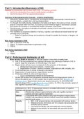
Part 1: Introduction/Summary of AD
Purpose: get familiarised with the basic idea of AD – no readings just facts
Use for the introduction if you need to briefly explain something
Use for the theory NOT for experimental evidence
Summary of Neurodegenerative Cascade – extreme simplification
Alzheimer’s Disease (AD) is a progressive neurological disorder pathologically characterised by
neuronal atrophy, synaptic loss, and neurodegeneration
It is thought that malfunctioning metabolism of amyloid-β will lead to the build-up of toxic amyloid
plaques composed within the brain. This build-up would therefore trigger a downstream cascade
that causes the formation of neurofibrillary tangles composed of tau protein
Simultaneously, microglia and astroglia are activated, causing chronic neuroinflammation and
neurodegeneration
This manifests as progressive deficits in memory, cognition, and behavioural impairments that will
ultimately lead to dementia
o Extent of cognitive change and symptoms is thought to parallel the formation of tangles, not
plaques
Main Genes Implicated in FAD
1. APP precursor of the Aβ
2. PSEN1 protease responsible for generation of Aβ
3. PSEN2
Main Genes Implicated in SAD
1. ApoE
2. TSEN2
Part 2: Pathological Hallmarks of AD
1. Brain atrophy (death of neurons) AD brain weighs 1/3 less than a healthy brain
a. One of the best established and validated biomarker and pathological hallmark of AD
used to stage the progression of AD pathology in the brain
b. Region where cell loss occurs corresponds to the symptoms of the disease
i. Widespread hippocampal atrophy = memory loss
ii. Degeneration of cortex = memory formation, organizational skills, empathy, etc\
c. Found dramatic loss of cortical cholinergic innervations
d. Degeneration of basal forebrain magnocellular neurons
e. Cholinergic deficits in cortex and hippocampus
i. Led to cholinesterase inhibitors as first treatment
f. Several MRI studies from laboratories worldwide reported that hippocampal volume in
clinical AD patients was 15–40% smaller than in healthy controls (HC) (Bosscher and
Scheltens, 2002). Hippocampal volume was already reduced by 15–30% at the mild
dementia stage of AD (van der Flier et al., 2005), and by 10–15% in the amnestic variant of
mild cognitive impairment (MCI) (Shi et al., 2009).
McDonald et al., 2012 hippocampal volume is correlated with severity of cognitive
disorders and episodic memory deficits
Aim Relationship between regional atrophy rates and cognitive decline in
patients with mild cognitive impairment
Method Longitudinal MRI scans analysed using high-throughput image analysis
Atrophy rats calculated by percent cortical volume loss between baseline
and 24 month scans
Results MCI left temporal atrophy rats associated with naming decline
Bilateral temporal, left frontal, and ACC atrophy rats associated with
semantic fluency decline
Left entorhinal atrophy rate associated with memory decline
Bilateral frontal atrophy rat associated with executive function
decline
Conclusio
, n
Critical Did not investigate this relationship in patients with AD – may not be
thinking able to be generalised
Sarazin et al., 2010
Aim Directly compared relationship with hippocampal atrophy and cognitive
decline in AD patients
Method
Results
Conclusio
n
Critical Hippocampal atrophy lacks specificity for AD
thinking
2. Extracellular deposition of B-pleated assemblies of AB peptide forming diffuse and neurite
senile plaques
a. No correlation between amount of amyloid in the brain and the severity of AD
b. More advanced the disease, the more widespread the amyloid plaques
c. Disrupt the neuropil and are activated glia, dystrophic axons, and dendrites
d. Identified through classic silver histochemical staining
e. Made up of amyloid Beta – amphipathic peptide that self-aggregates in the extracellular
space to form large spheres disrupting neuropil and destroying local synaptic fields
f. Viewed as critical indicator of AD because rare autosomal dominant forms of AD are
genetically linked to mutations in APP that is the precursor of abeta, one of the enzymes that
releases AB into the interstitial space
g. Aggregation propensities of AB modulated in two ways:
i. Directly
1. Pathogenic substitution present in the peptide transforms the mutant peptides
into a more aggregation-prone species
ii. Indirectly
1. Substitution promotes generation of longer AB peptides by modulating the
gamma-secretase proteolysis of APPC99
3. Intracellular neurofibrillary tangles (composed of tau protein) AKA NFTs
a. Abnormally phosphorylated (hyper) tau assembles into paired helical filaments (PHF) that
aggregate into neurofibrillary tangles (NFT) in the neuronal perikaryal and dystrophic
neurites
b. Identified through immunostaining and cresyl violet counterstaining
c. Occurs in the same population of neurons that are lost
d. Associated with some degree of glial reaction and synaptic loss in terminal zones
4. Cerebral amyloid angiopathy (CAA)
a. Where a beta becomes deposited in the arteries and the veins
5. Neuroinflammation
6. Increase in classic autophagosomes
a. Feature a double membrane that encapsulates heterogenous intracellular components seen
in AD brains
b. Deletion of key macroautophagy components like ATG5 or ATG7 in neurons in mice cause
motor deficits, ubiquitinated neuronal inclusions, and neurodegeneration
c. Abnormalities in autophagy are linked to tau pathology because neurons affected by loss of
function of NPC1 or NPC2 genes (Niemann-Pick’s disease type C) display tau inclusions
indistinguishable from those in AD
Discrepancy Between Pathology and Clinical Symptoms
Some individuals had fewer (or no) clinical symptoms despite increasing pathological change
Preservation of cognition in face of neuropathological lesions due to cognitive reserve
Brain resilience? differing extents of neurodegeneration as consequence of accumulation of
tangles and plaques
Diagnosing Pathological Hallmarks
1. Early diagnosis
a. Low levels of AB42 in the CSF
, b. Levels of phosphorylated tau higher than controls
c. MRI discloses regional brain atrophy in AD patients
d. Rates of atrophy
e. PET using SPECT shows decreased glucose utilization + early reduction of regional blood
flow in parietal and temporal lobes
f. Marked retention of brain penetrant tracers that bind to Aβ in areas of the brain that
commonly accumulate amyloid
The Problem with Diagnosis
1. Dementia occurs AFTER neuropathological changes in AD
a. Easy to make a diagnosis of dementia but it is a clinical challenge to determine AD at an
early stage
b. Neuropathological changes accumulate in the brain 10-15 years before the first sign of
clinical impairment
c. However, dementia due to AD is the last stage of disease
d. Diagnosing and treating AD at an early stage is thought to have much better therapeutic
success
i. ADD EVIDENCE HERE
e. We would need to develop a biomarker related to the pathological changes in AD or utilize
new imaging approaches to detect early stage AD
2. Limitations of current diagnosis methods
a. Using neuropsychological evaluation, patient interview, blood-sample analysis, and imaging
b. No useful biomarkers of disease stage
i. Has been one developed for atrophy but not all AD patients have atrophy in the early
stage of the disease
3. High right of undiagnoses
a. Variability of symptoms – dementia can be confused with other conditions like anxiety,
depression, sleep disorders, alcohol abuse
b. Onset of dementia is gradual which means that some patients (when in patient interviews)
may deny any cognitive deficits
c. How much of memory decline is considered normal ageing and how much is considered AD
– while standardization of diagnosis is attempted, it has yet to be achieved
Current Methods Under Investigation
1. Biomarkers: extremely helpful in not only confirming diagnosis to eliminate ambiguity, but it would
allow for the detection of early stage AD
i. Proteopathic tau seeding has been found to predict tauopathy (Holmes et al., 2014)
https://www.pnas.org/content/pnas/111/41/E4376.full.pdf
1. Purpose: reliably and specifically detects aberrant tau (not normal monomeric HD and
control) in AD brains SUPPLEMENT TO CURRENT AD AND TAUOPATHY-RELATED
POSTMORTEM DIAGNOSTICS
2. Note: seed = aggregates
a. Combined specificity of tau monoclonal biosensor cell line + sensitivity of FRET
flow cytometry to directly compare onset and progression of disease pathology
between seeding and histological deposition
b. Tau seeding increases in age-dependent manner in P301S mice and onset occurs
over 4 weeks before earliest histological marker of tau deposition
c. Tau seeding activity is an early and robust marker of taupathy
d. Transcellular propagation of protein aggregations (proteopathic seeds) drive
progression of neurodegenerative diseases in prion-like manner
e. Tau seeds propagate pathology through brain via cell-cell transfer in neural
networks
f. Unknown whether seeding anticipates and correlates with subsequent
development of pathology
g. FRET-based flow cytometry biosensor assay detected that proteopathic tau
seeding is an early and robust marker of tauopathy proximal role of tau seeds in
neurodegeneration
h. Seeding assay can overcome some limitations associated with standard
histological methods of AD diagnostics
i. Straightforward experimental setup
The benefits of buying summaries with Stuvia:
Guaranteed quality through customer reviews
Stuvia customers have reviewed more than 700,000 summaries. This how you know that you are buying the best documents.
Quick and easy check-out
You can quickly pay through credit card or Stuvia-credit for the summaries. There is no membership needed.
Focus on what matters
Your fellow students write the study notes themselves, which is why the documents are always reliable and up-to-date. This ensures you quickly get to the core!
Frequently asked questions
What do I get when I buy this document?
You get a PDF, available immediately after your purchase. The purchased document is accessible anytime, anywhere and indefinitely through your profile.
Satisfaction guarantee: how does it work?
Our satisfaction guarantee ensures that you always find a study document that suits you well. You fill out a form, and our customer service team takes care of the rest.
Who am I buying these notes from?
Stuvia is a marketplace, so you are not buying this document from us, but from seller denh11323. Stuvia facilitates payment to the seller.
Will I be stuck with a subscription?
No, you only buy these notes for $11.51. You're not tied to anything after your purchase.