Breast Pathology, UTSHC Fall 2022 D2 Pathology
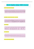
Breast Pathology, UTSHC Fall 2022 D2 Pathology What are the Distinguishing Features of the Breast From Other organs? - Nutritional support of another individual, as the major function - Marked changes of the structure throughout life - Having a social, cultural, and personal significance Normal Breast Structure and Histology - Two major structures: ducts and lobules - Lobule as the functional unit of the breast - Two types of epithelial cells (luminal and myoepithelial) - Two types of stroma (intralobular and interlobular) - Six to 10 major ducts orifices open onto the skin surface at the nipple what are the two major structures of the breast? ducts and lobules what is the functional unit of the breast? lobule what are the two types of epithelial cells that make up breasts? luminal and interlobular about how many major ducts orifices open onto the skin surface at the nipple? 6-10 What are some cells you will see in the Normal Breast Structure and Histology? - Intralobular stroma - Ducts - Lobules - Adipose tissue Life Cycle changes in breast tissue: after menarche Expansion of the lobular system and periodic remodeling during adulthood Life Cycle changes in breast tissue: onset of pregnancy Complete maturation and functional of the breasts with increase in number and the size of the lobules: permanent changes Life Cycle changes in breast tissue: after the third decade until menopause Involution and regression of the lobules and their specialized stroma and conversion of the interlobular stroma to adipose tissue Clinical Presentation of Breast Disease - Inflammation - Palpable mass - Lumpiness or other symptoms - Pain - Nipple discharge are the clinical presentation of breast disease good indicators of breast cancer? No, at most 7% turn out to be cancer what is the best way to check for breast cancer? mammogram screening, followed by palpable mass Clinical Presentation of the Breast Disease: Pain (mastalgia or mastodynia) - A common symptom. Maybe cyclic or noncyclic - Diffuse cyclic due to premenstrual edema - Localized noncyclic may be caused by ruptured cysts, physical injury, and infections - Almost all painful masses are benign - 10% of breast cancers presents with pain Clinical Presentation of the Breast Disease: Inflammation - Causes edematous and erythematous breast, mostly by infections, during lactation and breastfeeding - Different from "inflammatory" breast carcinoma Clinical Presentation of the Breast Disease: Nipple discharge - May be normal when small in quantity and bilateral - Papilloma arising in the large ducts below the nipple as the most common benign lesion producing a nipple discharge - Galactorrhea, not associated with malignancy - Increase in risk of malignancy in a woman with nipple discharge with age (carcinoma in 7% of women younger than 60 but in 30% of older women) Clinical Presentation of the Breast Disease: Lumpiness - A diffuse nodularity throughout the breast, a result of normal glandular tissue - If pronounced, imaging studies may be helpful Clinical Presentation of the Breast Disease: Palpable masses - Due to epithelial or stroma proliferation - Generally detected when are 2-3 cm in size - Round/oval circumscribed; 95 % are benign - Masses with irregular border as malignant - All palpable masses require evaluation Clinical Presentation of the Breast Disease: Gynecomastia The only common breast symptom in males, resulting from imbalance between estrogens and androgens Mammographic Screening Introduced in the 1980s as a means to detect small, nonpalpable, asymptomatic breast carcinomas what is the most common means to detect breast cancer? Mammographic Screening How do the sensitivity and specificity of mammography change with age? Why? increase with age, due to the changes in the breast tissue what are the probabilities of a mammographic lesion to be cancer at age 40 and over 50? 10% at 40, 25% over 50 what are the principle mammographic signs of breast carcinomas? densities and calcifications Ultrasonography in breast imaging distinguishes between solid and cystic lesions and defines more precisely the borders of solid lesions Magnetic resonance imaging (MRI) in breast imaging detects cancers by the rapid uptake of contrast agents due to i
Written for
- Institution
- Breast Pathology, UTSHC Fall 2022 D2 Pathology
- Course
- Breast Pathology, UTSHC Fall 2022 D2 Pathology
Document information
- Uploaded on
- December 6, 2022
- Number of pages
- 7
- Written in
- 2022/2023
- Type
- Exam (elaborations)
- Contains
- Questions & answers
Subjects
- breast pathology
-
utshc fall 2022 d2 pathology what are the distinguishing features of the breast from other organs nutritional support of another individual
-
as the major function marked change