TITLE Protein X-ray crystallography DATE
Reason for using X-ray crystallography:
Light microscope provide images of objects around
same size of wavelength of light (500nm)
but average size of a protein is -10nm and
an atom is 0.1 nm
:X-rays are used (wavelength 0.1nm(
=
X-ray microscopes are not feasible to make ... use crystallography
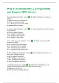
Crystallography process
- - -
I.
grow crystal 2.Put in front of X-ray 3. Electron density 4. build protein structure
beam - get diffraction map according map
pattern
AtOM:
nucleus + electrons
↳ v. Small:volume of atom is defined by electron cloud
molecule:
a set of bonded atoms
Crystal:
an ordered array of the same molecule in 3 dimensions
*
Only a
highly purified protein w/ all the same molecules can crystallize
to workout molecular structure, an
X-ray diffraction pattern is needed:
interaction of x-rays with electrons in crystal
https://bynikkib.com
,TITLE DATE
X-ray: a
travelling electromagnetic wave
↳ has electric and magnetic fields at
right angles to each other
Electric field: a
way of describing the electrostatic
force felt by a charged particle due to the
presence/motion of other charged particles
only consider this as it can interact w/ electrons
tells us which will move
way a the
charge
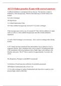
Electrons will scatter (or diffract) X-rays
by a single electron:
side view tOD View
W
In all
directions
by 2 electrons in close proximity:
each orin the structure becomes
a source of x-ray
detector observes scattering pattern
I resultant of scattered waves
added up
diffraction pattern depends on
structure
X-ray scattering by 2 electrons is like the 2-slit experiment:
↳ diffraction of water through gives
2 slits an
unvarying pattern
of peaks and troughs
depends on slit structure (e.g. width)
https://bynikkib.com
, TITLE DATE
Real structure of a whole protein gives a v. complex
diffraction pattern
:knowing how waves add up (like in 2-slit
experiment) will help deduce structure
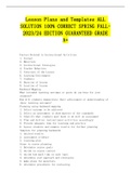
WAVES
the wave motion is cyclic
sin and cosine waves are horizontal and vertical projections
of a rotating line (aka radius (
the phase angle (0) Indicates the rotated position of
the line (radius)
O is measured:
where 1800: i radians
:I complete cycle 13000): phase angle of 20:25 radians
wave properties
1. Phase shift is
cyclic: crest at
every interval of 24 (2π, kπ, 64) and
trough at every odd i (π, 34, 5T (
2. Amplitude: maximum displacement from x axis
3.
Wavelength: distance between 2 peaks
https://bynikkib.com
Reason for using X-ray crystallography:
Light microscope provide images of objects around
same size of wavelength of light (500nm)
but average size of a protein is -10nm and
an atom is 0.1 nm
:X-rays are used (wavelength 0.1nm(
=
X-ray microscopes are not feasible to make ... use crystallography
Crystallography process
- - -
I.
grow crystal 2.Put in front of X-ray 3. Electron density 4. build protein structure
beam - get diffraction map according map
pattern
AtOM:
nucleus + electrons
↳ v. Small:volume of atom is defined by electron cloud
molecule:
a set of bonded atoms
Crystal:
an ordered array of the same molecule in 3 dimensions
*
Only a
highly purified protein w/ all the same molecules can crystallize
to workout molecular structure, an
X-ray diffraction pattern is needed:
interaction of x-rays with electrons in crystal
https://bynikkib.com
,TITLE DATE
X-ray: a
travelling electromagnetic wave
↳ has electric and magnetic fields at
right angles to each other
Electric field: a
way of describing the electrostatic
force felt by a charged particle due to the
presence/motion of other charged particles
only consider this as it can interact w/ electrons
tells us which will move
way a the
charge
Electrons will scatter (or diffract) X-rays
by a single electron:
side view tOD View
W
In all
directions
by 2 electrons in close proximity:
each orin the structure becomes
a source of x-ray
detector observes scattering pattern
I resultant of scattered waves
added up
diffraction pattern depends on
structure
X-ray scattering by 2 electrons is like the 2-slit experiment:
↳ diffraction of water through gives
2 slits an
unvarying pattern
of peaks and troughs
depends on slit structure (e.g. width)
https://bynikkib.com
, TITLE DATE
Real structure of a whole protein gives a v. complex
diffraction pattern
:knowing how waves add up (like in 2-slit
experiment) will help deduce structure
WAVES
the wave motion is cyclic
sin and cosine waves are horizontal and vertical projections
of a rotating line (aka radius (
the phase angle (0) Indicates the rotated position of
the line (radius)
O is measured:
where 1800: i radians
:I complete cycle 13000): phase angle of 20:25 radians
wave properties
1. Phase shift is
cyclic: crest at
every interval of 24 (2π, kπ, 64) and
trough at every odd i (π, 34, 5T (
2. Amplitude: maximum displacement from x axis
3.
Wavelength: distance between 2 peaks
https://bynikkib.com