Samenvatting neurophysiology
Book cellular and molecular neurophysiology
Lacture 1: Chapter 1,2,3
Ramon y Cajal was the first was the first who proposed that neurons are isolated units and that their
ends are not connected to each other. Terminal arborizations of the neurons are free and not connected
to other terminal arborizations.
Neurons are 1: excitable cells meaning these cells can receive information and translate it in electric
current. 2: secretory cells the secretory product is called neurotransmitters, release into the extracellular
space occurs only in restricted regions. 3: quiescent cells when the
neurons are lesioned they can not be replaced because they are
postmitotic cells, meaning they are not longer capable of under going
mitisis. However, they have the capacity to renew their
constituents(onderdelen) during their entire life, involving the precise
targeting of mRNA’s and proteins to particular cytoplasmic domain or
membrane areas. In humans, most neurons cannot divide after birth
since they are postmitotic cells. The large size of the nucleolus indicates
a high level of protein synthesis in the neurons.
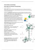
Golgi´s staining the cells are fixed and stained by potassium dichromate
and silver nitrate. Biocytin can also be used to
visualise neurons( see figure on the left)
Three functions of the nerve cell:
Metabolic function : synthesis and degradation of the
proteins is in cell body, requires anterograde and
retrograde transport
Signal reception : dendrites and soma receive
information via postsynaptic
regions(neurotransmitter receptors)
Secretory functions: in axon terminals the
presynaptic regions are located( synaptic vehicles
containing neurotransmitters, synthesis of enzymes,
volted- dependent channels)
Soma has a couple functions: 1. Soma is the place of
the synthesis of many macromolecules that are
required for the structure and the function of the
neuron 2. Soma receives synaptic contacts from
other neurons.
,Axon differ from the dendrites for example: 1. axons have an uniform diameter along its entire extent,
while the diameter of the dendrites decreases with branching. 2. Axons do not contain ribosomes of
endoplasmic reticulum, while dendrites do. Almost all parts of the cell body are rich of ER but the axon
hillock is not. 3.Another difference is the orientation of the microtubules. Axons have uniform polarity of
the microtubules, meaning that al plus/ ends are pointing away from the cell body. Conversaly, dendrites
have antiparallel microtubules.
Some neurons are termed ´spiny´ because they have between 40 000-100 000 spines on their
dendrites( vb pyramidal neuron in the cerebral cortex, purkinje cell of the cerebellar cortex), others are
termed ‘smooth’ with only a few spines on the surface of the dendrite( neurons of the pallidal complex).
Dendrites and soma receive numerous synaptic contacts from other neurons and constitute the main
receptive area of neurons. In response to afferent information, the neurons generate electrical signals
such as postsynaptic potentials (excitatory =EPSP and inhibitory= IPSP) in the postsynaptic membrane
and integrate the afferent information. These postsynaptic potentials propagate and summate in the
somatodendritic compartment, then they propagate to the axon where they generate the action
potential. At the beginning of the axon(a) there is an initiation of the action potential this place is called
the axonal initial segment and the beginning of the axon is termed the axon hillock. On the inside, axon
exists of specialized bundle of microtubules, while on the outside it is covered by myelin sheath. Myelin
is formed by glial cells and is needed for the conduction of the action potential. The length of the axon
depends on its function and the place in the brain. The axons end in a terminal arborization where the
vesicles with neurotransmitters can be released. Another type of axon terminal is the ´boutons an
passant´ described as a swelling on the axon that makes a
non-terminal synaptic contact with another neuron but is not
the ending of an axon. The main characteristic of axons is
their capacity to open trigger the sodium action potentials
and to propagate them over considerable distance without
any decrease in amplitude. When the action potentials reach
the axon terminals these trigger calcium action potentials
which may cause the release of the neurotransmitters
contained in the axon terminals, the synaptic vesicles. Certain
regions, such as the initial segment, Nodes of Ranvier and axon terminals can also be receptive areas.
On the inside of the axons and dendrites there are
microtubules which transport particles to the positive side
in the cell. Wherefor do we need microtubules and do the
axons only transport to the axon terminal and the dendrites
both sides?(p 10)
Figure on the right demonstrates anterograde axonal
transport. On the top a neuron is illustrated, than a ligature
is placed on the axon. The third axon shows an
enlargement in the diameter near the ligature. In the fourth
axon the ligature is removed and the diameter of the axon
in that place decreases. These experiment of Weiss and
Hiscoe(1948) confirmes anterograde transport in the axon
,nerves of the chicken and suggest the existence of two
types of transport. Anterograde(kinesin richting+) –
from cell body to the terminals, retrograde(dynein
richting -)- from the terminals to the cell body.
Another study showed that kinesin mutations impair
the function impair the function of action potential
and the release of vesicles at the nerve terminal. Thus
kinesin appears to be required for axonal transport:
for example axonal transport of NA+ channels to the
Ranvier Nodes and the Ca+ channels to the
presynaptic membrane. The function of the
retrograde transport is the removal of the misfolded
or aggregated proteins back to the cell centre for
recycling or degradation.
Convergence of information the information is split in
multiple paths to control for example multiple
muscles. Divergence of information the information of
multiple neurons comes into one. Anterograde
inhibition(feedforward inhibition) a neuron inhibits
another neuron by the activation of an inhibitory
interneuron. Recurrent inhibition( feedback
inhibition) a neuron inhibits itself by a recurrent
collateral of its own axon which synapses on a
inhibitory neuron. See figure on the left.
What are the respective functions of the inhibitory
local circuit neuron and Renshaw’s cell?
After the activation of the muscle the neuron need to inhibit itself. This happends via the recurrent axon
collateral and the Renshaw’s cell otherwise there wil be a countineous activation of the muscle.
Chapter 2
There are roughly twice as many glial cells as there are neurons in the central nervous system. Glia cells
can be classified on their anatomical position: 1. Central glia are found in the central nervous system,
compare 4 cell types: astrocytes,oligodendrocytes, microglia( these three types together are also called
interstitial glia, because they are found in the interneural space) and ependymal cells wich form the
epithelial surface covering the walls of the cerebral ventricles and of the central canal of the spinal cord.
2. Peripheral glia comprise a single type: Schwann cells, these cells ensheeath the axons and
encapsulate the cell bodies of neurons.
, 3 characteristics that distinguishes glial cells from the neurons:1. They do not generate or conduct an
action potential. 2. They do not establish chemical synapses between themselves, or any other cells. 3.
Glial cells are capable of division far at least several years postnatally.
Astrocytes are small strar-shaped cells. There are two kinds of astrocytes recognized some of them
contain in their cytoplasm glial filaments and those are located in the white matter, while others contain
a few glial filaments and are located in the gray metter.
What do the astrocytes: 1. Maintain the blood-brain barriere in the adult brain The essential
characteristic of astrocytic processes is their termination on the walls of blood vessels in astrocytic end
feet. Here the end feet are joined by gap junction and desmosomes, forming a ‘palisade’ between
neurons and vascular endothelial cells. Astrocytes by themselves are not the blood-brain bariere, but the
epithelial cells which are are joined toghether by the tight junctions are. However, astrocyte cells play an
important role in maintanance of the development of the blood-brain barriere. 2. Regulate the ionic
composition of the extracellular fluid Astrocytes play a role in controlling the composition of the
extracellular fluid, for exaple the potassium. The extracellular potassium needs to be tightly regulated
otherwise the increase of potassium could cause a depolarization of neurons. The astrocytes regulate the
extracellular potassium( in particular near the nodes of Ranvier) through a mechanism of ‘spatial
buffering´. This means the astrocyte take up potasium ions in regions where the concentration rises and
releases through their end feet into the blood vessels or vicinity(nighbourhood). 3. Regulate efficacy of
synaptic transmission After neurotransmitters are released during synaptic transmission, they need to
be removed from the extracellular space to prevent the extracellular neurotransmitter concentration
from raising( long lasting activation of the receptors> could damage the neurons) Astrocytes not only
play a role in the reuptake of neurotransmitters but are also involved in the synthesis of it. Astrocytes
release gliotransmitters when the intracellular Ca2+ concentration is getting high.( why?)’
What are the difference in function of Schwann cell and the oligodendrocytes? Are they both involved
in myelination?
Is it that Schwann cells are involved in myelination in the Periferal nervous system and oligodendrocytes
in the Central nervous system
Oligodendrocytes are involved in forming the meyelin sheath, to isolate the axons , induce the
formation of the NA+ channels at the nodes of Ranvier, to allow the fast propogation of Na+ action
potential. Meyelin consists of 70 procent fat and 30 procent protein.
Neuron/glia cells: astrocyte, microglia
Microglia : phagocytes, release mediators to influence the neuronal excitability, regulation adult
neurogenesis.
Node of Ranvier is the place where the sodium channels are concentrated.
Chapter 3
The benefits of buying summaries with Stuvia:
Guaranteed quality through customer reviews
Stuvia customers have reviewed more than 700,000 summaries. This how you know that you are buying the best documents.
Quick and easy check-out
You can quickly pay through credit card or Stuvia-credit for the summaries. There is no membership needed.
Focus on what matters
Your fellow students write the study notes themselves, which is why the documents are always reliable and up-to-date. This ensures you quickly get to the core!
Frequently asked questions
What do I get when I buy this document?
You get a PDF, available immediately after your purchase. The purchased document is accessible anytime, anywhere and indefinitely through your profile.
Satisfaction guarantee: how does it work?
Our satisfaction guarantee ensures that you always find a study document that suits you well. You fill out a form, and our customer service team takes care of the rest.
Who am I buying these notes from?
Stuvia is a marketplace, so you are not buying this document from us, but from seller valeriyag. Stuvia facilitates payment to the seller.
Will I be stuck with a subscription?
No, you only buy these notes for $7.36. You're not tied to anything after your purchase.