Kidney Overview
Notice: To optimize your learning in this course, we advise that you complete the labs and modules as indicated in the BIOD 152 Lab Schedule.
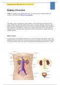
The urinary system is composed of a pair of kidneys, each with one ureter which leads to the urinary bladder from which the urethra carries...
1
Kidney Overview
Notice: To optimize your learning in this course, we advise that you complete the labs and
modules as indicated in the BIOD 152 Lab Schedule.
The urinary system is composed of a pair of kidneys, each with one ureter which leads to the
urinary bladder from which the urethra carries wastes as urine from the body. The ureters and
urethra are tubes which transport the urine from kidney to bladder and bladder to the outside the
body respectively. The bladder is a urine storage container. The kidneys serve many functions
among which are: fluid homeostasis, filtration of wastes and homeostasis of blood volume and
chemical makeup.
Kidney Anatomy
An adult kidney is bean-shaped, about the size of a can of soup and weighs about 5 ounces. The
right kidney (because of the liver's location just above it) lies slightly lower than the left and both
are protected by the rib cage since they are located in the lumbar region between the T-12 and L-
,Portage Learning / BIOD 152 Kidney module 5 exam
2
3 vertebra. The kidney's medial surface is concave and has a cleft called the renal hilus leading to
a space within the kidney called the renal sinus. The ureters, blood vessels, and nerves are
located in the sinus and entering the kidney at the hilus. On top of each kidney is an adrenal
gland.
The kidney surface is protected by three layers of specialized tissue. The renal capsule is a tough
fibrous outer skin of the kidney which protects it from injury and infection. Outside of the renal
capsule is a fatty layer that protects the kidney from trauma which is called the adipose capsule.
The outer renal fascia is dense fibrous connective tissue which keeps the kidney in place inside
the abdominal cavity.
The three separate regions evident in a vertical section of the kidney are the cortex, the medulla,
and the pelvis. The outer renal cortex, just inside the renal capsule, is a continuous outer region
with a number of projections (cortical columns) that extend down between the renal medulla
pyramids. Located within the cortex are the glomerular capsule and the distal and convoluted
tubule sections of the nephrons along with associated blood vessels.
,Portage Learning / BIOD 152 Kidney module 5 exam
3
Deeper within the kidney lies the renal medulla which is divided into sections called pyramids
that point toward the center of the kidney. Located within the medulla are the Loop of Henle and
the collecting duct sections of the nephrons along with associated blood vessels.
The centermost section of the kidney near the renal hilus is the renal pelvis which constitutes a
funnel-shaped tube that connects to the ureter as it leaves the hilus. Several extensions of the
pelvis called calyces collect urine which drains continuously into the renal pelvis and
subsequently into the ureter, which transports the urine to the bladder to be stored.
Blood and Nerve Supply
The kidneys are innervated by many blood vessels so that they can filter the blood to regulate its
composition with the renal arteries delivering about 1200 ml of blood per minute of blood
directly from the abdominal aorta which amounts to 20% of the cardiac output. The renal arteries
branch into 5 segmental arteries which divide further into lobar arteries then further into
The benefits of buying summaries with Stuvia:
Guaranteed quality through customer reviews
Stuvia customers have reviewed more than 700,000 summaries. This how you know that you are buying the best documents.
Quick and easy check-out
You can quickly pay through credit card or Stuvia-credit for the summaries. There is no membership needed.
Focus on what matters
Your fellow students write the study notes themselves, which is why the documents are always reliable and up-to-date. This ensures you quickly get to the core!
Frequently asked questions
What do I get when I buy this document?
You get a PDF, available immediately after your purchase. The purchased document is accessible anytime, anywhere and indefinitely through your profile.
Satisfaction guarantee: how does it work?
Our satisfaction guarantee ensures that you always find a study document that suits you well. You fill out a form, and our customer service team takes care of the rest.
Who am I buying these notes from?
Stuvia is a marketplace, so you are not buying this document from us, but from seller THEEXCELLENCELIBRARY. Stuvia facilitates payment to the seller.
Will I be stuck with a subscription?
No, you only buy these notes for $37.99. You're not tied to anything after your purchase.