Summary
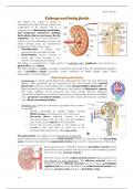
Summary Kidneys and body fluids
- Course
- Institution
Unlock the secrets of the kidneys and body fluids with this comprehensive guide! Learn about the kidney's anatomy, remarkable filtration process, its regulation of body fluid volume and composition, and its role in maintaining plasma osmolarity. Discover how the kidneys handle toxins and essential ...
[Show more]