OEDEMA, ELECTROLYTE BALANCE, AND DIABETES INSIPIDUS
OEDEMA
BODY FLUIDS AND HAEMODYNAMIC
BODY FLUIDS
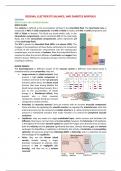
The oedema is defined as the accumulation of fluid in the interstitial fluid. The total body mass is
composed of 40% of solid components and 60% of fluids in males, and 45% of solid components and
55% of fluids in females. The fluids are found within cells, in the
intracellular compartment, which constitutes 2/3 of the total body
fluids, and in the extracellular compartment, which represents 1/3
of the total body fluids.
The ECF is present as interstitial fluid (80%) and plasma (20%). The
changes in the equilibrium of these fluids, particularly the movement
of fluids of the intravascular compartment into the extravascular
compartment, are the cause of oedema. Note that interstitial fluid is
required to allow the correct diffusion of nutrients and oxygen from
blood to tissues. Without it, all plasma will pass into the interstitium
resulting in oedema.
BLOOD VESSELS
The haemodynamic in different vessels of the vascular system is different. Each blood vessel is
characterised by some properties; they are.
• Larger arteries (or elastic arteries): they
present a high elastic component to
maintain and resist to the high pressure
presents within them; the elastic recoil
ensures that even during diastole the
blood moves along blood vessels; this is
due to the accumulation of elastic
energy (i. e. Windkessel effect); they
present also a thick muscular
component, despite being less relevant
compared to smaller arteries.
• Arterioles (or muscular arteries): they are arteries with an increase muscular component;
these arterioles by regulating the smooth muscles can regulate the arteriole tone; note that
arterial pressures can be measured in smaller arteries (e. g. radial, brachial arteries) since the
pressure is maintained constant; their major function is to control the peripheral vascular
resistance.
• Capillaries: they are made of a single endothelial layer, which ensures and facilitates the
diffusion; they have a narrow lumen and cross section; however, the total area of all capillaries
is the highest of the entire vascular system (as alveoli for lungs); since the total area increases,
both the velocity and the pressure significantly decrease, thereby allowing gas and nutrient
exchange; the bloodflow in capillaries is dictated by the activity of arterioles and precapillary
sphincter; the pressure drops from 94mmHg (MAP) to 30mmHg, and it is important to avoid
the breakage of the capillaries.
• Veins: they are the thinner blood
vessels, despite having a larger
diameter compared to arteries; their
pressure is low to negligible (10-
0mmHg), and they act as a volume
reservoir.
, CAPILLARIES
The capillaries have a diameter of 8-10m (erythrocytes 5-8m, cardiomyocytes 20-25m and length
of 100m), and the endothelial membrane is made of cytoplasmic protrusions. The exchange along
capillaries may occur through endothelial junctions for fluids and
electrolytes, or via caveolae (transcytosis). The innermost part of
the endothelial layer is made of glycocalyx. This layer helps to
maintain the endothelial integrity and impermeability. The
glycocalyx is made of glycated proteins and therefore it is
negatively charged; it pushes away proteins, thus lowering the
permeability to plasma proteins. Hence, while glucose (MW
180g/mol) can pass, the albumin (negatively charged, MW 66kDa)
cannot. Therefore, the high-MW molecules cannot pass through this wall, as well as the negatively
charged ones.
STERLING FORCES
The capillaries exchange is dictated by four main forces, which are also named Sterling forces, which
are:
• Capillary hydrostatic pressure (Pc): it is the pressure
that moves fluids from the capillary to the
interstitium; it is of 30mmHg at the arterial end, while
of 10mmHg at the venous end.
• Capillary osmotic pressure (c): it is the pressure
exerted by the plasma proteins (i. e. albumin) to
move fluids from the interstitium to the capillaries; it
is of about 28mmHg.
• Interstitial hydrostatic pressure (Pi): it is the pressure
that moves fluids from the interstitium to capillaries;
it is of about 3mmHg.
• Interstitial capillary pressure (i): it is the pressure
exerted by interstitial proteins to move fluids from capillary to the interstitium; it is of about
8mmHg.
These different forces can be put together in the Starling law, which states that fluid movements
between blood and tissues are determined by differences in hydrostatic and oncotic pressures
between the plasma and interstitial fluid, combined with the endothelial permeability. The formula
is:
𝑭𝒍𝒐𝒘 = (𝑷𝒄 − 𝑷𝒊 ) − 𝝈(𝝅𝒄 − 𝝅𝒊 ) = 𝝈 × 𝑵𝑭𝑷
The is the reflection coefficient, and it describes how much the capillaries are permeable. If it is
equal to 1, it means capillaries are reflecting everything and their permeability is at its maximum
number (i. e. they are impermeable). Conversely, if is lower than 1 and tend to 0, the capillaries are
very permeable (inflammation). When capillaries increase their permeability to plasma proteins, the
oncotic pressures are compromised, resulting in an increase of the I and in a decrease of c.
The algebraic sum of the Sterling forces determines the flow of fluids in the interstitium. At the arterial
end, the net filtration pressure (NFP) is of +7mmHg, which means that the fluid moves from capillaries
to the interstitium. This is called filtration. Conversely, the net filtration pressure at the venous end is
of -6mmHg, which means that the fluid moves from the interstitium to capillaries. This is called
reabsorption.
LYMPHATIC CIRCULATION AND TYPES OF OEDEMAS
LYMPHATIC COMPENSATION
The volume of fluid that is filtered into the interstitium from capillaries in the arterial end within an
hour is of 1200mL. In the venous end, instead the reabsorption is of about 1080mL/h. The small
The benefits of buying summaries with Stuvia:
Guaranteed quality through customer reviews
Stuvia customers have reviewed more than 700,000 summaries. This how you know that you are buying the best documents.
Quick and easy check-out
You can quickly pay through credit card or Stuvia-credit for the summaries. There is no membership needed.
Focus on what matters
Your fellow students write the study notes themselves, which is why the documents are always reliable and up-to-date. This ensures you quickly get to the core!
Frequently asked questions
What do I get when I buy this document?
You get a PDF, available immediately after your purchase. The purchased document is accessible anytime, anywhere and indefinitely through your profile.
Satisfaction guarantee: how does it work?
Our satisfaction guarantee ensures that you always find a study document that suits you well. You fill out a form, and our customer service team takes care of the rest.
Who am I buying these notes from?
Stuvia is a marketplace, so you are not buying this document from us, but from seller marcocassina02. Stuvia facilitates payment to the seller.
Will I be stuck with a subscription?
No, you only buy these notes for $6.32. You're not tied to anything after your purchase.