Notes of respiratory pathophysiology.
Physiology of respiratory systems and determinants of respiration.
Obstructive pathologies: asthma, chronic bronchitis, pulmonary emphysema, and COPD.
Restrictive pathologies: interstitial lung diseases (ILDs) and idiopathic pulmonary fibrosis (IPF).
Pneumo...
RESPIRATORY PATHOPHYSIOLOGY
DETERMINANTS OF RESPIRATORY FUNCTION
RESPIRATORY PHYSIOLOGY
THREE RESPIRATORY DETERMINANTS
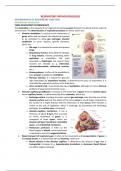
The respiration is the capacity of our organism to exchange gases between the blood and the external
environment. The determinants of respiratory function are three, which are:
• Alveolar ventilation: it consists in the movement of
gases within and out alveoli; the alveoli are required
to be ventilated to allow gas exchange; alveolar
ventilation to occur requires specific elements,
which are:
Rib cage: it is essential to sustain and protect
lungs.
Respiratory muscles: they allow the changes
in lung volume, thereby promoting either
expiration or inspiration; the most
important is diaphragm, but several other
muscles are involved (e. g. intercostal,
sternocleidomastoid, abdominal muscles,
etc.).
Airway patency: it refers to the possibility to
have airways available for ventilation.
Nervous stimulus: it is required to give the
right stimulation to respiratory muscles; it determines the pace of respiration; it is
controlled by respiratory centres in the brainstem.
Air O2 content of air: it can dictate the rate of ventilation, although it is not an intrinsic
problem of lungs (environmental issue).
• Alveolar-capillary gas diffusion: it consists in the movement of gases from the alveolar space
to the capillary lumen; it is determined by three elements, which are:
Exchange surface: it is large to ensure correct gas exchange; note that the sum of the
alveolar surface given by the alveoli of the same acinus is much larger compared to
the surface of a single alveolus with the dimension of that acinus; their function is
similar to the one of capillaries, which is exchange; by promoting and facilitating
exchange, the surface area is increased.
Alveolar-capillary membrane: it has a
thickness of about 0.2m, thus ensuring
an easier movement of gases; it is
composed of three layers, which are
alveolar cell type I membrane, interstitial
space, and endothelial membrane.
Pulmonary perfusion: it refers to the
blood supply of alveoli; correct perfusion
is required to correctly oxygenate blood.
• Blood transport of respiratory gas: it refers to the movement and transportation of gases in
the blood vessels; it is determined by two elements, which are:
Blood velocity in the alveolar capillary: it determines the amount of gas that is
transported; increases (e. g. hyperdynamic circulation) or decreases (heart failure)
can affect oxygen exchange.
, O2 transport in the blood: it is related to mechanisms that are used to transport
oxygen in the blood (i. e. haemoglobin); defects in its transport can be caused by
anaemia.
RESPIRATORY SYSTEM AND POLLUTANTS
The respiratory system is the part of the body that is mostly exposed to the external environment.
The air contents can easily enter within the respiratory system. However, large particles (>5m) are
filtered by hairs and mucous present in the nostrils; smaller
particles (3-5m) tend to stop in the bronchial system due to
the presence of the mucociliary escalator, while the smallest
particles (<3m) may reach the alveoli.
These smallest particles can be taken up by alveolar
macrophages, which have the function of phagocyting and
degrading them. There are some particles that are difficult to
digest, or that cause macrophages worn out (i. e. frustrated
macrophages). An example is asbestos. Other possible
pollutants are PM10, PM5, and PM2.5, among which PM2.5 are the most dangerous.
LUNG VOLUMES AND CAPACITIES
The measurements of lung volumes can be assessed during spirometry, and they are divided into 4
volumes and 4 capacities, which are:
• Inspiratory reserve volume (IRV): it is the volume
of air that can be inspired over and above the
normal tidal level with a maximum effort; it is of
about 3000mL.
• Expiratory reserve volume (ERV): it is the volume
of air that can be expired with maximum effect
after the end of a normal tidal expiration; it is of
about 1200mL.
• Residual volume (RV): it is the volume of air that
remains in lungs after the most forceful expiration; it is of about 1200mL.
• Tidal volume (TD): it is the volume of air that is either expired or inhaled during each normal
breath; it is of about 500mL.
• Vital capacity (VC): it is the volume of air that is expired with a maximum effort after a
maximal inspiration (ERV+TD+IRV); it is of about 4700mL.
• Functional residual capacity (FRC): it is the volume of air that remains in lungs after normal
expiration (ERV+RV); usually, it is taken as main parameter to start respiratory tests, and it is
of about 2400mL.
• Inspiratory capacity (IC): it is the volume of air that can be inspired from normal expiratory
levels (IRV+TD); it is of about 3500mL.
• Total lung capacity (TLC): it is the volume of air that can be contained by lungs (VC+RV); it is
of about 5900mL.
Other important parameters are used in the dynamic spirometry, in
which the flow of air is measured, rather than the lung volumes; they
are:
• Fraction of expired volume in the first second (FEV1): it is the
maximal volume that is exhaled at the end of second 1 from the
beginning of an expiration that follows a forced inspiration; it is
reduced in obstructive conditions (e. g. asthma).
• Forced vital capacity (FVC): it refers to the volume of air that can
be exhaled after the deepest inhalation.
BRONCHIECTASIS AND PULMONARY ATELECTASIS
The bronchiectasis is a pathological condition that is characterised by an abnormal and irreversible
dilation of the bronchi. It can be either localised or diffused, and it is a consequence of inflammation
of the structural components of the bronchial wall. It can be caused by
bacteria, such as P. aeruginosa, H. influenzae, and S. aureus, which
secrete proteases and other toxins that damage the respiratory
epithelium and prevent mucociliary clearance.
The pulmonary atelectasis is another respiratory disorder characterised
by a loss of air content within alveolar spaces. It can be caused by three
main events, which are:
• Compression of pulmonary parenchyma: it can
be caused neoplasms and cysts.
• Extrapulmonary compression: it can be caused
by pleural effusion.
• Bronchial obstruction: it can be caused by
foreign bodies and neoplasms.
DYSFUNCTIONS ON ELEMENTS OF THE THREE DETERMINANTS
HYPOVENTILATION
The hypoventilation refers to a reduction of gas exchange between the atmosphere and alveolar air.
It is a problem related to alveolar ventilation, and it can be caused by problems affecting its main
elements, which are:
• Damaged rib cage: it is associated with bone
injuries, which impair respiratory mechanics
since it ensures lung expansion during
inspiration (e. g. trauma, rib fractures,
crushing, etc.).
• Problems in respiratory muscles: it can be
associated with myopathies, either congenital
(e. g. DMD, ALS, etc.) or acquired (e. g.
poliovirus, iron lung), and with paralysis of
respiratory muscles; the failure of respiratory
muscles will cause failure in ventilation.
• Airways without patency: it is associated with obstruction (e. g. inhaled object) and
bronchoconstriction; the latter is caused by the contraction of smooth muscles of the
bronchioles, and the most common associated disorder is asthma.
• Neurological syndromes: they are associated with a decrease nervous stimulation; they affect
the respiratory centres of the brainstem and nerve conduction; examples are poliovirus, ALS,
autoimmune conditions, and opioid overdose.
• O2 reduction in air: it is associated with decrease oxygen intake; it is observed at high altitudes
and in indoor spaces.
The benefits of buying summaries with Stuvia:
Guaranteed quality through customer reviews
Stuvia customers have reviewed more than 700,000 summaries. This how you know that you are buying the best documents.
Quick and easy check-out
You can quickly pay through credit card or Stuvia-credit for the summaries. There is no membership needed.
Focus on what matters
Your fellow students write the study notes themselves, which is why the documents are always reliable and up-to-date. This ensures you quickly get to the core!
Frequently asked questions
What do I get when I buy this document?
You get a PDF, available immediately after your purchase. The purchased document is accessible anytime, anywhere and indefinitely through your profile.
Satisfaction guarantee: how does it work?
Our satisfaction guarantee ensures that you always find a study document that suits you well. You fill out a form, and our customer service team takes care of the rest.
Who am I buying these notes from?
Stuvia is a marketplace, so you are not buying this document from us, but from seller marcocassina02. Stuvia facilitates payment to the seller.
Will I be stuck with a subscription?
No, you only buy these notes for $7.47. You're not tied to anything after your purchase.