Module
6:
The
Respiratory
System
Anatomy
and
Mechanics
Main
Roles
of
the
Respiratory
System:
1.
Oxygen
Intake
and
Carbon
Dioxide
Removal
:
facilitates
oxidative
metabolism
by
taking
in
oxygen
and
expelling
carbon
dioxide
2.
pH
Regulation:
plays
a
critical
role
in
maintaining
body
pH
by
managing
cardon
dioxide
levels
3.
Immune
Defense
:
the
respiratory
system,
with
its
vast
surface
area
exposed
to
about
10,000
liters
of
air
daily,
is
a
significant
site
for
pathogen
defense.
→
Flow
(F)
=
pressure
gradient
(
△
P)/resistance
®
-
R
is
related
to
the
radius
of
the
tube
-
1/r^4
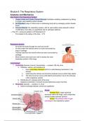
Respiratory
System:
-
Air
enters
the
body
through
the
nose
and
mouth
-
Air
then
enters
the
trachea
which
is
a
tube
surrounded
by
rings
of
cartilage
-
Trachea
branches
into
bronchi
which
are
also
surrounded
by
cartilage
-
Brochi
branch
more
and
more
until
it
reaches
the
main
respiratory
portion
of
the
lungs
Lung
Airways:
1.
Conducting
(trachea,
bronchi,
bronchioles)
→
contains
150
mls
of
air
a.
Cleanses,
warms,
and
moistens
air
i.
Uses
mucociliary
transport
which
is
a
self-clearing
mechanism
in
the
airways
ii.
Cells
lining
the
trachea
and
bronchis
produces
mucus
which
traps
debris
iii.
The
cilia
move
the
mucus
(with
debris)
and
pushes
it
up
to
the
entryway
of
the
respiratory
system
b.
Distributes
air
to
deeper
parts
of
the
lung
c.
Dialation/constriction
of
smooth
muscle
determines
ease
of
air
flow
2.
Respiratory
(alveoli)
→
contains
3
L
of
air
a.
Gases
exchange
between
alveoli
and
capillaries
Alveoli:
-
Alveolar
Sacs
:
larger
spherical
structures
within
the
lungs,
each
comprising
several
smaller
spheres
(alveoli)
-
Alveoli
:
the
primary
sites
of
gas
exchange
in
the
respiratory
system -
Inside
of
the
alveoli
(where
the
air
is
present)
has
a
massive
area
for
gas
exchange
(~70
square
meters
in
total)
which
is
essential
for
efficient
oxygen
intake/
CO2
expulsion
-
The
outside
is
covered
with
capillaries
which
are
also
crucial
for
transporting
gases
Type
I
Alveolar
Cells:
These
cells
form
the
majority
of
the
alveolar
surface
and
are
extremely
thin
and
flat.
This
minimal
thickness
facilitates
a
short
diffusion
distance
for
gases
between
the
air
in
the
alveoli
and
the
blood
in
the
capillaries
.
Type
II
Alveolar
Cells
:
Less
numerous
and
cover
less
surface
area
and
have
two
functions:
1.
Stem
Cells
:
they
can
regenerate
alveolar
cells,
which
is
crucial
for
lung
repair
and
maintenance
2.
Surfactant
Production
:
They
produce
a
substance
called
surfactant
that
reduces
surface
tension
within
the
alveoli
,
preventing
them
from
collapsing
during
exhalation
and
making
the
process
of
inhalation
more
efficient.
Ventilation
and
Lung
Mechanics:
Negative
Pressure
Breathing
→
the
way
we
get
air
into
our
lungs
-
This
type
of
breathing
is
based
on
creating
a
pressure
gradient
between
the
lungs
and
the
atmosphere
-
The
diaphragm
(a
dome-shaped
skeletal
muscle
located
below
the
lungs)
is
central
to
this
process.
When
you
inhale,
the
diaphragm
contracts
and
flattens
which
increases
the
thoracic
cavity’s
volume
.
This
expansion
reduces
the
pressure
inside
the
cavity
below
that
of
the
outside
atmosphere,
causing
air
to
flow
into
the
lungs.
Muscle
Actions
During
Breathing:
1.
Inhalation
:
a.
Normal
Conditions:
Primarily
involves
the
diaphragm.
As
it
contracts
and
moves
downward,
it
enlarges
the
chest
cavity
and
decreases
lung
pressure,
pulling
air
in.
b.
Active
Conditions
:
During
exercise
or
deep
breathing,
additional
muscles
assist
in
increasing
lung
volume.
These
include
the
muscles
of
the
rib
cage,
which
lift
the
ribs
outward
and
upward,
further
enlarging
the
thoracic
cavity.
2.
Exhalation
:
a.
Passive
Exhalation
:
Typically
occurs
during
restful
breathing.
When
the
diaphragm
relaxes,
it
moves
back
to
its
dome
shape,
decreasing
the
volume
of
the
thoracic
cavity.
The
lungs
also
contract
due
to
their
elastic
properties,
pushing
air
out.
This
process
does
not
generally
require
effort
and
is
driven
by
the
natural
recoil
of
the
lungs
and
the
relaxation
of
the
diaphragm.
→
The
lungs
possess
natural
elasticity
,
meaning
after
being
stretched
during
inhalation,
they
tend
to
return
to
their
original
size.
This
elastic
recoil
helps
in
passive
exhalation,
simplifying
the
breathing
process
under
normal
conditions
without
requiring
muscular
effort
b.
Active
Exhalation
:
During
vigorous
activities
or
rapid
breathing,
exhalation
becomes
an
active
process
involving
the
abdominal
muscles
and
the
internal intercostal
muscles
(those
between
the
ribs).
These
muscles
help
compress
the
thoracic
cavity,
forcefully
expelling
air
from
the
lungs.
Importance
of
the
Diaphragm
and
Other
Respiratory
Muscles:
-
Diaphragm
:
Essential
for
normal,
quiet
breathing
and
acts
as
the
primary
muscle
for
inhalation.
-
Rib
Cage
Muscles
:
Important
during
deep
or
forceful
breathing,
assisting
in
expanding
the
thoracic
cavity
more
than
what
can
be
achieved
by
the
diaphragm
alone.
-
Abdominal
and
Intercostal
Muscles
:
Crucial
during
active
exhalation,
aiding
in
quickly
expelling
air
from
the
lungs.
Boyle’s
Law:
This
law
states
that
at
constant
temperature,
the
pressure
of
a
gas
is
inversely
proportional
to
its
volume.
In
the
context
of
breathing,
when
the
diaphragm
contracts
and
flattens,
it
increases
the
volume
of
the
thoracic
(chest)
cavity,
which,
according
to
Boyle's
Law,
decreases
the
pressure
inside
the
lungs.
Ventilation
Cycle:
1.
Inhalation
:
initiated
by
diaphragm
contraction,
expanding
the
thoracic
cavity
and
reducing
lung
pressure
2.
Breathing
Pause
:
After
inhalation,
there's
often
a
brief
pause
where
the
pressures
between
the
alveoli
and
the
atmosphere
equilibrate,
causing
no
air
flow
(zero
flow
state).
3.
Exhalation
:
Typically
passive
in
relaxed
states,
where
the
diaphragm
relaxes,
the
thoracic
cavity's
volume
decreases,
pressure
inside
the
lungs
increases,
and
air
is
pushed
out.
This
is
facilitated
by
the
elastic
recoil
of
the
lungs,
which
inherently
want
to
return
to
a
smaller
size.
Tidal
Volume
(TV):
the
amount
of
air
inhaled/exhaled
in
a
normal
breath
(~0.5
L)
Minute
Ventilation:
Calculated
by
multiplying
the
tidal
volume
by
the
breathing
frequency
(number
of
breaths
per
minute).
→
Ex
:
breathing
0.5
liters
per
breath,
10
times
a
minute,
results
in
a
minute
ventilation
of
5
liters.
Adaptations
in
Breathing
During
Activity:
-
Increased
Ventilation
Needs
:
During
intense
activities
like
exercise,
the
body
requires
increased
air
(and
oxygen)
intake,
which
can
increase
the
minute
ventilation
up
to
20
times
the
normal
resting
state.
Blood
flow
to
the
respiratory
system
can
also
increase
up
to
three
times
to
support
this
heightened
activity.
-
Active
Exhalation
:
Unlike
passive
exhalation
during
rest,
active
exhalation
is
necessary
during
vigorous
activity.
This
requires
engaging
additional
muscles,
like
the
abdominal
muscles
and
internal
intercostal
muscles,
to
forcibly
expel
air
from
the
lungs
quickly,
allowing
for
faster
subsequent
inhalations.
Key
Concepts: